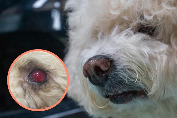
There are many conditions which could cause a dog's eye to become red. Looking at the specific symptoms can help us to determine the cause. When there is inflammation, redness in the sclera (white of the eye) or the conjunctiva is red can suggest various infections and ailments. However, when blood appears to be in the eye of the dog, it suggests a condition known as hyphema. This is when blood accumulates in the anterior chamber of the eyeball, i.e. the space between the cornea and the iris. It has specific causes which need to be determined before treatment can commence.
If you see there is blood in your dog's eye, it is important to know why. AnimalWised helps explain by providing the causes and treatment of hyphema in dogs.
What is hyphema in dogs?
The anterior chamber of the eyeball is the space between the cornea and the surface of the iris. The anterior chamber houses the aqueous humor, the clear fluid which covers the eye. Extravasation of blood from the anterior uvea (iris and ciliary body) can leak into the anterior chamber. The components of the blood (cells and blood plasma) mix with the aqueous humor, causing it to acquire a reddish color.
It is important to distinguish this between other types of eye redness. When hyphema in dogs occurs, the eye will look glassy red due to the blood cells mixed with the normally transparent aqueous humor. It is usually seen in the lower part of the eye, as the blood content falls due to gravity. However, when the dog moves its head, the blood is distributed throughout the anterior chamber, showing a homogeneous reddish coloration.
Blood in the anterior chamber does not usually clot easily due to the release of fibrinolysins (enzymes that dissolve fibrin clots) from the iris. For this reason, blood clots do not usually appear until 4-7 days after the start of bleeding.
Depending on the severity and extent of the condition, ocular hyphema can be classified into:
- Grade I: when it occupies less than a third of the anterior chamber.
- Grade II: when it occupies half of the anterior chamber.
- Grade III: when it occupies three-quarters of the anterior chamber.
- Grade IV: when it occupies the entire anterior chamber.
In addition to the grades of hyphema, it is crucial to monitor for other symptoms that may arise, such as changes in the dog's behavior or vision difficulties, which can indicate the severity and impact on the dog's overall health. If you want to know more about issues dogs can have with their eyes, take a look at our article on common eye problems in dogs.
Causes of hyphema in dogs
Hyphema in dogs can be due to both localized and systemic causes. We break these two broad categories down into individual causes:
Localized causes of hyphema in dogs
There are several eye disorders that can trigger hyphema in dogs:
- Trauma to the eyes: it is the most frequent cause. Trauma due to a blow, fall, collision or any other reason can lead to the extravasation of blood into the eye.
- Uveitis: inflammatory process that affects the uvea (the vascular tunic of the eye)
- Glaucoma: caused by damage to the optic nerve or retina, often leading to increased intraocular pressure. Find out more with our article on glaucoma in dogs.
- Retinal detachment: more common in senior dogs, it has various causes which can be genetic or acquired.
- Ocular neoplasms: such as lymphomas. Lymphoma in dogs is a serious disease which can be fatal, but hyphema is likely only one symptom.
- Congenital eye abnormalities: genetic inheritance which may be related to certain breeds, but can affect any dog.
Generally, localized hyphema are usually unilateral. This means they only affect one eye, e.g. if they receive trauma to one eye and not the other.
Systemic causes of hyphema in dogs
The two main systemic causes that can cause hyphema in dogs are:
- Hypertension: increased blood pressure which can itself be due to various problems. These include heart disease, hormonal imbalance and even stress. Learn more with our related article on congestive heart failure in dogs.
- Coagulation disorders: thrombocytopenia, coagulopathies such as Von Willebrand disease, anticoagulant poisoning, ehrlichiosis, etc.
Systemic hyphema is usually bilateral, i.e. it affects both eyeballs. Additionally, it's essential to address any underlying health issues promptly, as untreated systemic conditions can lead to more severe complications beyond hyphema.
Symptoms of hyphema in dogs
Although most of us will be alerted to the presence of hyphema in dogs due to the appearance of blood in the dog's eye, there are other signs which we might notice. The symptoms of hyphema in dogs are usually:
- Reddish band in the anterior chamber of the eye: it will be more or less extensive depending on the severity of the condition. When the animal moves their head, the blood is dispersed throughout the chamber, showing a homogeneous reddish coloration. When at rest, the blood in the dog's eye will be more concentrated in the lower section.
- Blepharospasm: closure of the eye, blinking and twitching due to eye pain.
- Epiphora: continuous tearing, especially when the cause is traumatic.
- Swelling or cloudiness: a sign of inflammation or additional complications in the eye, which may require immediate attention to prevent further damage.
- Changes in vision: the dog might demonstrate difficulties navigating familiar environments, bumping into objects, or showing reluctance to move in bright light.
Monitoring these symptoms closely and communicating them to a veterinarian can help in determining the stage of hyphema and any necessary interventions.
Diagnosis of hyphema in dogs
The diagnosis of hyphema in dogs should be carried out by a veterinarian who specializes in ophthalmology. Specifically, the diagnosis of hyphema in dogs includes:
- Complete ophthalmological examination: the anterior chamber of the eye must be studied from a lateral perspective using a slit light. This is to correctly locate the hemorrhagic focus. In case of unilateral hyphema, it is important to perform a complete examination of the healthy eye, as it can provide useful information for the diagnosis.
- Tonometry: to measure intraocular pressure.
- Ocular ultrasound: a very useful diagnostic tool that also helps to establish a better prognosis of the condition.
- Blood tests and blood pressure measurement: it is essential in case of bilateral hyphema, since the cause is usually systemic.
To reach the definitive diagnosis of ocular hyphema, it is necessary to rule out the following differential diagnoses:
- Hemorrhagic inflammatory exudate in the anterior chamber: unlike hyphema, the hemorrhagic inflammatory exudate has a more yellowish color, a denser appearance and occupies the entire anterior chamber (not just the lower part).
- Hemorrhage in the vitreous chamber: in this case the reddish coloration is behind the pupil (in the vitreous chamber). However, the possibility of concomitant hyphema and vitreous hemorrhage should not be ruled out. In the case of vitreous hemorrhage, examination of the fundus part of the eye will be difficult or impossible.
- Hemorrhage in the cornea or iris: observing the anterior chamber from a lateral perspective, it is possible to differentiate the precise location of the hemorrhagic focus.
Accurate diagnosis is critical in determining the appropriate treatment plan and preventing potential complications that could affect the dog's vision and overall health.
Treatment of hyphema in dogs
Hyphema is considered an ophthalmologic emergency that requires immediate medical attention. When you suspect that dog may be suffering an intraocular hemorrhage, do not hesitate to go to a veterinary clinic as soon as possible. The veterinary team will most likely stabilize the ophthalmological emergency in situ, and then refer the case to an ophthalmology specialist.
Treatment of hyphema in dogs should include:
- Topical or systemic anti-inflammatories: corticosteroids are generally used, since NSAIDs (Non-Steroidal Anti-Inflammatory Drugs) such as ibuprofen for dogs are discouraged due to their possible antiplatelet effect.
- Mydriatic or cyclopegic: these are drugs that cause the pupil to dilate and are used in cases of hyphema to prevent the appearance of synechiae (adhesions). Tropicamide can be used in mild hyphemas (grade I or II) or Phenylephrine 10% in severe hyphemas (grade III and IV). It should be taken into account that the use of these drugs is controversial, since they can predispose to an increase in intraocular pressure.
- Topical carbonic anhydrase inhibitors: such as dorzolamide or brinzolamide. They will only be used in cases of ocular hypertension.
- Analgesics: opioids such as buprenorphine can be used when there is eye pain, especially in traumatic cases.
- Rest: recuperation is important, so the dog may be kept under observation in a veterinary hospital.
When the hemorrhage becomes a blood clot, it is advisable to perform an intracameral injection of TPA (Tissue Plasminogen Activator). Occasionally, surgical removal of the clot may be necessary. These procedures should only be performed by specialist ophthalmic surgeons. Additionally, follow-up visits are crucial to monitor the healing process and adjust treatments as needed to ensure the best possible recovery for the dog.

Prognosis and complications of hyphema in dogs
The prognosis of hyphema in dogs depends on its underlying cause and its eventual progression:
- Grade I: usually resolves in less than a week.
- Grades II and III: can take several weeks to resolve.
- Grade IV: often causes atrophy and shrinking of the eyeball, also known as phthisis bulbi. The dog will be blind in this eye as it will not longer function.
Typically, the blood lodged in the anterior chamber gradually drains through the iridocorneal angle. However, sometimes intraocular hemorrhage can lead to the following complications:
- Formation of intraocular synechiae: adhesions can form between the clot and the cornea or between the clot and the iris.
- Ocular hypertension (values above 25 mmHg) and glaucoma.
- Cataracts: often seen when the dog has cloudy eyes, possibly with blood also.
- Retinal detachment.
- Retinal degeneration.
- Atrophy of the eyeball or phthisis bulbi.
- Blindness: when the eye damage is extensive, the dog will no longer be able to see. This is worse if the condition is bilateral as it affects both eyes. If you suspect this has happened to your dog, take a look at our article on how to know if your dog is blind.
Early detection and treatment are paramount in improving the outcome for dogs with hyphema, potentially reducing the risk of long-term complications and preserving vision. With ocular diseases, trauma and other conditions, various symptoms can appear. To learn more, take a look at our articles on why a dog has blue eyes and the causes of green ocular discharge in dogs.
This article is purely informative. AnimalWised does not have the authority to prescribe any veterinary treatment or create a diagnosis. We invite you to take your pet to the veterinarian if they are suffering from any condition or pain.
If you want to read similar articles to Blood In My Dog's Eye, we recommend you visit our Eye problems category.
- Huguet, E., & Díaz, C. (2013). Chapter 1: Hyphema. In Red Eye. Ophthalmology in colors. Editorial Multimédica.
- Maggs, D. G., Miller, P. E., & Ofri, R. (2008). Slatter's fundamentals of veterinary ophthalmology. 4th ed. Saunders Elsiever.
- Morales, I., Gutiérrez, E., De León, M., Ferrer, O., & Corbera, JA (2006). Veterinary ophthalmology. The use of tissue plasminogen activator (TPA). Magazine of the Spanish Veterinary Collegiate Organization, 14-19