Seromas are an accumulation of serous fluid in the subdermal area under the skin, although sometimes they can also develop between the musculature. They are one possible complication of various surgeries, especially after ventral midline surgery. Although many are naturally reabsorbed into the dog's body, in some cases it may be necessary to extract the liquid or even insert a drainage device.
To prevent their appearance, precision surgery and careful closure of the incision will reduce the underlying causes of seroma. Keep reading this article from AnimalWised to learn more about seroma in dogs, specifically looking at symptoms, post-surgical treatment and prevention.
What is a seroma in dogs?
A seroma is defined as the accumulation of serous fluid, specifically blood plasma and inflammatory fluid, outside the blood vessels. It accumulates under the skin in the subdermal area. It differs from hematoma in that the seroma lacks red blood cells.
Canine seromas can also appear in other locations, including:
- Shoulders
- Ears
- Neck
- Head
- Brain
A canine seroma is a soft and generally painless mass, occurring in empty spaces in the fatty layer located between the skin and the muscles. It may appear as the result of a blow or incision, such as you would find in common canine surgeries. This is a result of the inflammatory process instigated by the dog's autoimmune defenses.
However, a seroma should not be confused with an abscess as it does not contain any pus. We explain the differences between the two in our article on abscesses in dogs.
The development of seromas can sometimes indicate an underlying issue with the healing process. It's important to monitor the site closely to ensure it does not develop into something more serious, such as an infection or other complications.
Causes of seroma in dogs
Seromas mainly occur after surgery as a type of surgical complication. They are especially common in abdominal surgeries in the ventral midline, including spaying and neutering operations. The risk of seroma formation in ventral midline surgery is approx. 10% (i.e. 1 in 10 dogs will develop a seroma after surgery).
This complication is more likely if, during the procedure, the veterinary surgeon has performed any of the following:
- Excessive dissection of the dog's subcutaneous tissue and skin
- Careless handling of tissues
- Poor closure of incisions
Other possible causes of seromas are alterations in blood clotting, punctures or trauma. Part of the reason they are relatively common is because spaying and neutering surgeries are common abdominal operations carried out on dogs. Since there are no red blood cells, we should differentiate seroma in dogs from other surgical complications. We discuss these further in our article on reasons why a dog bleeds after spaying.
In addition to surgical factors, certain breeds may have a predisposition to developing seromas due to their genetic makeup or body structure. Understanding these factors can help in planning preventive measures during surgery.

Symptoms of seroma in dogs
Seromas in dogs cause a fluid-filled swelling under the skin. If the dog has had surgery, the seroma will be found around the site of incision. Most often they occur subdermally, but they may occasionally occur between the muscular layers. The serous fluid is not blood, but we shouldn't be able to see the fluid color under the skin. The mass will feel soft due to the fluid accumulation, not hard as occurs with many skin tumors on dogs.
Generally the dog may present the following clinical signs associated with seroma:
- Swelling of the area (possibly accompanied by pain)
- Reddening of the skin
- An increase in temperature around the wound
- Clear fluid leaking from the incision
- Infection
Depending on the location of non-surgical seromas, the dog may show neurological signs of distress. These can include seizures and coma in cases where seromas develop in the brain. If it is a cervical seroma, it may cause discomfort and reduce neck mobility. If they occur on the shoulders, it may cause them pain when walking and other mobility issues.
In some cases, the dog might exhibit behavioral changes such as increased restlessness or reluctance to engage in usual activities. Monitoring these signs can provide valuable insights into the severity of the seroma.
Diagnosis of seroma in dogs
If a lump or swelling appears close to the surgical wound in the days following surgery, this is a reason to suspect seroma. Different to seromas, hematomas and hernias are caused by the reopening of the wound, especially in cases of abdominal surgery.
An ultrasound can make this distinction and can recognize if the lump contains organs or blood serum. Seromas are also different from hematomas as the fluid can be extracted by a needle. In the case of cranial seroma, advanced imaging techniques such as magnetic resonance imaging or computed tomography should be used.
Moreover, a fine needle aspiration may be performed to extract the serous fluid, which can then be analyzed to confirm the absence of red blood cells, solidifying the seroma diagnosis. This is a critical step to ensure appropriate treatment is administered.
Canine seroma treatment
In most dogs, no treatment may be required. This is because the seroma is often reabsorbed by the skin in about 10-20 days. In other cases, the following steps can be taken:
- Extraction: if the fluid cannot be completely reabsorbed due to size or severity, a needle may can be used for extraction.
- Drainage: in the most severe cases, it may be necessary to temporarily place a drain so that serous fluid does not continue to accumulate in the area. A drain is a tube that passes through the skin, connecting with the seroma, allowing fluids to escape. Drainage can be passive in nature, with the application of a compress. Active drainage using suction can be used for the most severe cases. In this case, the drain should not be removed until the fluid flow is no greater than 0.2 ml/kg per hour.
- Corticosteroids or surgery: encapsulation of the seroma can occur if left untreated. When the seroma hardens, it will leave an unattractive scar and corticosteroids, or even surgery, may be necessary.
- Antibiotics: the seroma can also become infected, causing an abscess and pus to leak from the scar. In these cases, antibiotics should be used to treat the bacterial infection.
- Analgesics: if the dog experiences pain or discomfort, analgesics or anti-inflammatory drugs should be administered.
It should be noted that while seromas often resolve on their own, consistent monitoring is crucial to prevent complications. Veterinarians might also recommend physical therapies to assist with fluid drainage and enhance healing.

Prevention of seroma in dogs
To prevent seroma formation, care should be taken during surgery and in the postoperative period:
- During surgery: tissue trauma and dissection should be minimized. Care should be taken to not leave any spaces during closure. This can be achieved by suturing the subcutaneous tissue to the underlying fascia to eliminate spaces. An effective suture pattern is the continuous quilting pattern, in which one in every three stitches is anchored to the fascia.
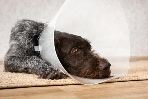
- During the postoperative period: dressings or compresses should be used. The dog should also be kept in a quiet place, given moderate rest and be given an Elizabethan collar to stop any licking of the area.
While seromas in dogs should not be ignored, they do not often pose a serious threat to the dog's health. It is not a reason to avoid neutering your dog, an intervention which provides many benefits to your dog, including increasing their life expectancy.
Additionally, ensuring the dog maintains a healthy weight and receives proper nutrition can support healing and reduce the risk of seroma development. Regular follow-ups with a veterinarian can also help in early detection and timely management of any complications.
This article is purely informative. AnimalWised does not have the authority to prescribe any veterinary treatment or create a diagnosis. We invite you to take your pet to the veterinarian if they are suffering from any condition or pain.
If you want to read similar articles to Seroma in Dogs - Post-Surgical Treatment, we recommend you visit our Other health problems category.
- Welch, T. (2019). Small animal surgery (5th edition). Elsevier.