Corneal Sequestrum in Cats


See files for Cats
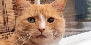
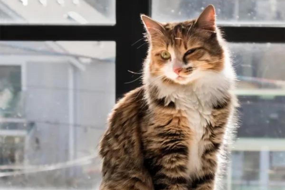
Cats have very distinctive eyes. They can be of varying colors, as well as appear in different sizes according to breed and other factors. While their pupils can dilate according to changes in light, they should generally have a similar appearance. When alterations to the eye occurs, it is a likely sign of a health problem. When you see there is a black spot on your cat's eye, it could appear as if it were a problem such as a corneal ulcer. However, it may also be a relative rare condition known as a corneal sequestrum.
At AnimalWised, we look at the causes and treatment of corneal sequestrum in cats. We look at the symptoms other than a black spot on the cat's eye, as well as any other important information.
What is corneal sequestrum in cats?
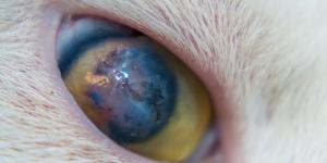
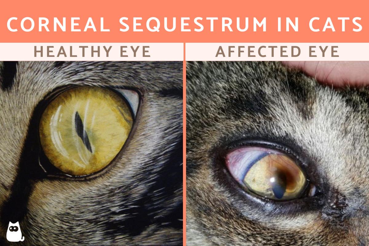
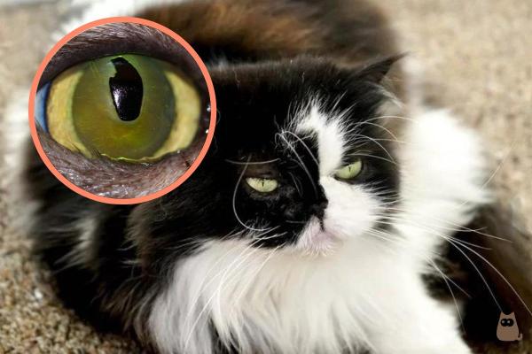
Also known as feline corneal degeneration, corneal sequestrum in cats is a condition of the cornea which causes focal degeneration of collagen. It results in the presence of porphyrins, a dark brown pigment. This pigment is diffusely located in the upper stroma of the cornea and gradually transforms into an irregular black plaque. This plaque appears as a black spot which is sometimes surrounded by new blood vessels. It penetrates the stroma of the cornea. This can lead to perforation and even the eventual loss of the affected eye.
In the initial stages it can be confused with a corneal ulcer, but the sequestrum evolves to a dark color. It does not stain when fluorescein dye is applied. This ocular disorder produces a lot of pain in our cats due to the degree of penetration into the cornea.
This corneal disorder appears mainly in cats between 2 and 7 years of age and usually only unilateral, i.e. it affects only one of the feline's two eyes. It is a condition which has a certain predisposition according to breed. The Persian cat appears to have greater prevalence to this condition, although it is also relatively more frequent in the following breeds:
- Siamese
- Sphynx
- Himalayan
- Common European
- Exotic Shorthair
Learn more about frequent medical issues for the Persian cat breed with the common diseases of Persian cats.

Symptoms of feline corneal sequestrum
The clinical signs that cats present with corneal sequestrum include the following:
- Black spot located centrally in the eye
- Eye pain
- Photophobia (intolerance to light)
- Epiphora (excessive tearing)
- Blepharospasm (excessive blinking/eye twitching)
- Mucopurulent discharge
- Corneal edema (fluid build up)
- Corneal neovascularization (new blood vessels appear)
- Cellular infiltrate in the cornea
- Protrusion of the nictitating membrane
In general, it can be suspected that a cat has corneal sequestrum when they have it has an ulcer that does not heal or changes color. We can see a black spot appearing on their cornea, as well as other symptoms such as the third eyelid showing. It will make the shape of the eye appear as if it has changed the black plaque is similar to the color of the cat's pupil.
As we stated above, many people confuse this with the presence of a corneal ulcer. Take a look at our article on corneal ulcers in cats to learn more.

Causes of feline corneal sequestrum
Now that we know the nature of sequestrum in cats and its symptoms, we are going to see its underlying causes. Scientific research on feline corneal sequestrum are not yet fully conclusive and have not fully established its cause. However, there does appear to be a link between this condition and continuous irritation of the cornea derived from processes such as:
- Entropion
- Corneal ulcers
- Trichiasis
- Alterations of the tear ducts
Feline corneal degeneration can also have a hereditary component, as can be seen in the fact it is more prevalent in certain breeds. It can be secondary to trauma and some authors suggest the cause may be primary stromal dystrophy.
Another cause that has been associated with feline corneal sequestrum is the feline herpesvirus type 1 infection (feline rhinotracheitis). This is because it is common for this virus to produce ocular symptoms such as ulcers or conjunctivitis, being isolated in up to 50% of cases of this condition.
Diagnosis of feline corneal sequestrum
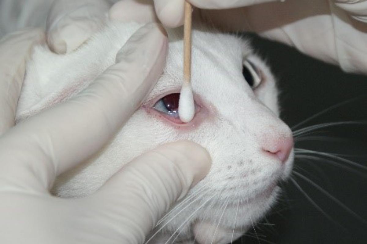
To diagnose corneal sequestrum in a cat, a full eye examination should be performed. This will begin with viewing the eye under white light to see the color of the plaque. It is seen when there is a dark spot appears which is more or less centered on the cornea. It is usually surrounded by new blood vessels. It will not appear when stained with fluorescein drops, so rose bengal will need to be used.
It is a good idea to also perform a Schirmer test to determine the amount of tears produced and the measurement of intraocular pressure with a tonometer. This will also be used to examine the fundus of the eye.
In order to diagnose a feline herpesvirus infection, you should:
- Take a sample of the conjunctiva
- Perform a PCR
- Use of optical coherence tomography (OCT)
Optical coherence tomography is a useful technique for the diagnosis of this condition and it does not require sedation of the cat as it does not contact the corneal surface. This technique consists of the emission of an infrared light source that penetrates the tissues of the eye and is reflected on the retina. When the reflection returns, the light creates an interference that produces a color image that shows the structures of the eye and their measurement. Colder colors indicate less thickness and warm colors indicate greater thickness.
This technique is used for diagnosis. It will also help inform the decision of which surgical treatment technique to use and for postoperative control to assess the continuity of the layers and the integration of the graft in the cornea.

Treatment of feline corneal sequestration
The treatment of feline corneal sequestrum is medical or surgical depending on the severity of the condition. In addition, the treatment will also be based on its underlying cause. Such treatment will range from the use of medication to surgical intervention to correct any damage to the eye.
Depending on the degree of pain and depth of the abduction, medical treatment will be carried out.
- In the mildest cases: consists of the use of antibiotic eye drops (frequently with tobramycin, chloramphenicol or ciprofloxacin), anti-inflammatories (prednisolone or dexamethasone) or ophthalmic ointments. This may be administered together with recombinant interferon 2alpha and antiviral treatment (idoxuridine, acyclovir, trifluorothymidine) in case of associated rhinotracheitis.
- In moderate to severe cases: surgical treatment will be necessary by performing techniques such as keratotomy, which consists of removing dead tissue so that the cornea can regenerate.
- In very severe cases: when the sequestrum is very deep, corneal grafts will be necessary to fill in the removed area. Other less-used techniques are corneal-conjunctival translation, flaps or corneal transplants.
This article is purely informative. AnimalWised does not have the authority to prescribe any veterinary treatment or create a diagnosis. We invite you to take your pet to the veterinarian if they are suffering from any condition or pain.
If you want to read similar articles to Corneal Sequestrum in Cats, we recommend you visit our Eye problems category.
- Stephen, J. (2022). Atlas of Clinical Ophthalmology of the Dog and Cat, 2nd Edition. Assisi Group Biomedia, S. L.

