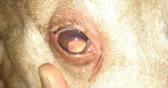
Lens Luxation in Dogs - Dislocated Eye Lens in Dogs


See files for Dogs
The ophthalmic diseases dogs can suffer from are varied and can affect different eye structures. Lens luxation is a displacement of the lens inside the eye due to a tear in the ligaments that hold the lens in suspension. Lens luxation is a common pathology that affects the lens of some dogs, particularly Collies, German Shepherds, and Shar-Pei. Some types of lens luxation are a medical emergency and require immediate ophthalmologic treatment. Therefore, it is important to know the signs that may be associated with this condition in order to take early action.
In the following AnimalWised article, you will learn everything you need to know about lens luxation in dogs, common symptoms, treatment, and surgery.
What is lens luxation in dogs?
Before explaining what lens luxation is, we should look at the structure of the eye to understand what this pathology consists of.
The lens works with the cornea to properly focus light onto the retina (a light-sensitive layer of tissue at the back of the eye). When light hits the retina, special cells called photoreceptors convert the light into electrical signals. Under normal conditions, the lens is located right in the center and behind the pupil, suspended by so-called zonula fibers or ribbons.
Lens dislocation occurs when the zonula fibers tear and the lens shifts from its normal anatomical position.
Weakness of the lens ligaments is known to be hereditary in terrier breeds, Chinese Shar Peis and Border Collies. If you have one of these dog breeds, it is important to watch for signs of discomfort or changes in the appearance of the eye and call your veterinarian immediately if you notice any changes.
Common eye problems in dogs usually require a visit to the veterinarian, as many of these conditions can lead to blindness if left untreated. For more information on the most common eye issues in dogs and their symptoms, see this other article on the 10 most common eye issues in dogs.
Types of lens luxation in dogs
Lens dislocations can be classified according to different criteria. Depending on whether the tear of the zonular fibers is complete or incomplete, one speaks of:
- Lens dislocation: When the zonular fibers tear completely, i.e. at an angle of 360º, there is a complete dislocation of the lens.
- Lens subluxation: When only part of the fibers break, there is a partial dislocation of the lens.
Depending on the position in which the lens falls into the orbit, we can divide the luxation into two different categories:
- Anterior luxation: in this case, the lens falls forward into the eye. Anterior luxations are considered an ophthalmic emergency because they block the outflow of fluid from the eye, leading to glaucoma or increased intraocular pressure. This is extremely painful and can lead to permanent blindness.
- Posterior luxation: In this case, the lens falls backwards into the eye and rarely causes discomfort.
Finally, we can also classify luxation in dogs into two categories, depending on the cause:
- Primary luxation: it is caused by a defect in the proteins that form the zonule. It occurs in young animals with a congenital weakness of the zonule or in older dogs due to a chronic degeneration of the zonule.
- Secondary luxation: Rupture of the zonule occurs as a result of a previous condition, such as trauma, ocular perforation, cataract, glaucoma, intraocular tumor, or uveitis.
Eye problems can be difficult to detect, but any visible change in your dog's eye structure should be investigated. For example, opacities are a common eye symptom that may indicate that the dog has cataracts or glaucoma. Continue reading this other article to learn more about the causes of cloudiness in your dog's eye.
Causes of lens luxation in dogs
The causes that may lead to dislocation or subluxation of the lens in dogs are the following:
- Genetic defect: There are certain breeds, especially terriers, Chinese Shar Peis, and Border Collies, that are born with a structural weakness of the zonula that causes the zonula to break at a certain moment and the lens to dislocate. These cases of luxation usually occur in young animals.
- Advanced age: With increasing age, chronic degeneration of the zonula may occur, leading to complete or partial rupture of the zonula.
- Other ocular diseases: Lens luxation is often secondary to other diseases such as uveitis, glaucoma, ocular cancer, ocular or head trauma, cataract, intraocular tumors or inflammation in the eye.
It is common for middle-aged to older dogs to develop a bluish, translucent haze in the lens of their eye. This is considered a normal age-related change in the lens. Read this other article to learn more about this condition in older dogs called nuclear sclerosis.
Symptoms of lens luxation in dogs
The clinical signs that may be observed in dogs with lens luxation are:
- Signs of ocular pain: lacrimation (epiphora), squinting or keeping the eye(s) closed, photophobia and depressed mood.
- Visual disturbances: Signs of visual impairment or blindness.
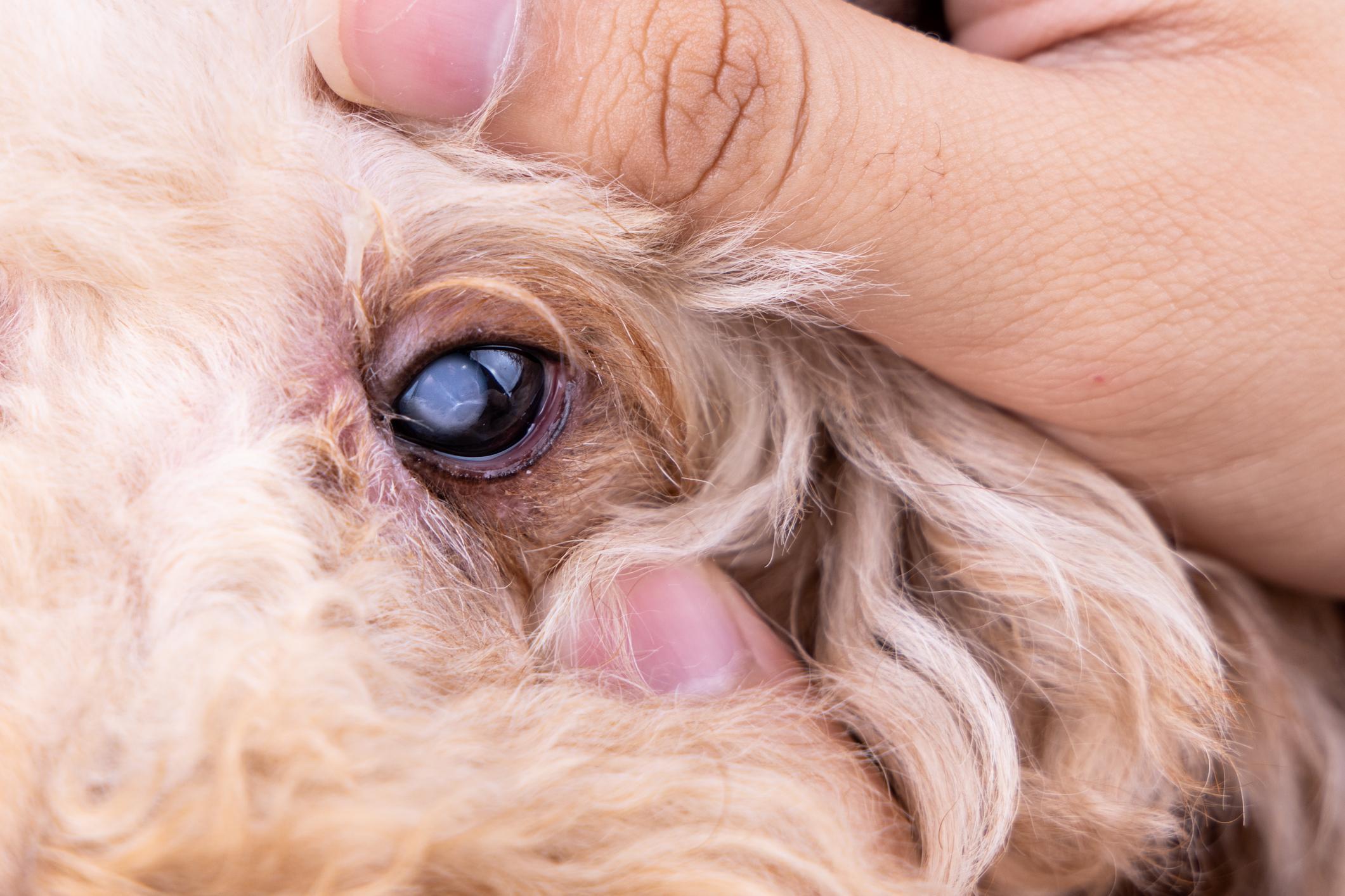
- Changes in the transparency of the eye: both changes in the transparency of the cornea (due to the appearance of corneal edema) and the lens itself (due to the development of a cataract in the displaced lens). This is also called opacification of the eye.
- Aphakic crescent: when the lens is displaced in relation to the center of the pupil, a crescent-shaped silhouette is formed. This sign is typical of lens subluxations.
- Iridonesis: This is abnormal movement or shaking of the iris (colored part of the eye).
- Lenticulodonesis: abnormal movements of the lens.
In addition, other complications associated with lens luxation can occur. The most common and important is the development of glaucoma in the affected eye. In these cases, scleral vessel blockage, corneal edema, pupil dilation (mydriasis), eye pain, and vision loss are often observed.

Diagnosis of lens luxation in dogs
Diagnosis of this pathology may be more or less straightforward depending on whether it is a subluxation, anterior or posterior dislocation. In any case, the diagnostic protocol may include the following steps:
- Ophthalmic examination: in the case of a lens subluxation, the aphakic crescent mentioned above is observed. In the case of a posterior dislocation, the retinal vessels can be seen with the naked eye (without the need for a posterior examination). In the case of an anterior dislocation, the lens is observed anterior to the iris. If the lens has developed a cataract, diagnosis is easier than if the lens is still transparent. For greater precision, the examination may need to be performed with a slit lamp.
- Ocular ultrasound: In cases where the diagnosis is complicated, it may be useful to perform an ocular ultrasound to determine the displacement of the lens more precisely.
Treatment and surgery of lens luxation in dogs
The treatment of this ocular pathology depends essentially on the type of dislocation diagnosed:
- Surgery: for anterior luxations, the best treatment in this situation is to remove the lens from the front of the eye by surgery. This procedure requires the use of the surgical microscope and specialized microsurgical skills. The chance that a patient will be able to see two years after surgery is about 50%. Your dog may remain in the hospital for a few days after surgery for careful monitoring, treatment and recovery. Until surgery can be performed, it is important to control pain and keep signs of glaucoma under control.
- Treatment of clinical symptoms: For posterior luxations, the lens is usually left in the vitreous and only therapy is initiated to relieve clinical signs and possible complications of the dislocation. However, most of the time this type of luxation causes little or no discomfort. It is possible to preserve patients' vision to varying degrees with drops that constrict the pupil. However, there is always the possibility that the luxated lens may come forward (anterior luxation) and become stuck when the pupil is dilated. Also, keep in mind that the medications used to constrict the pupil are not suitable for every patient. Therefore, the patient should be carefully observed for several hours after the first application before starting long-term treatment.
- Treatment of primary pathologies: For secondary dislocations, it is important to determine treatment for the primary pathology that caused the dislocation in the first place, as it may be a disease that can also affect the other eye.
The goal of these treatments is to relieve pain (in the case of an anterior luxation) and restore vision as much as possible. If the lens has been dislocated for some time, the chance of restoring vision is reduced. In fact, most luxations are considered emergencies and must be treated immediately (within 48 hours) or the animal could become permanently blind. With timely treatment, vision can be reasonably good in most cases, although it will never be perfect.
When it comes to lens luxation, time is of the essence. Therefore, you should see your veterinarian immediately if you suspect that your pet is suffering from an eye condition.
Glaucoma has devastating effects that can lead to blindness in your pet. Read this other article on glaucoma in dogs to learn more about the symptoms and treatment for this common eye disease.
This article is purely informative. AnimalWised does not have the authority to prescribe any veterinary treatment or create a diagnosis. We invite you to take your pet to the veterinarian if they are suffering from any condition or pain.
If you want to read similar articles to Lens Luxation in Dogs - Dislocated Eye Lens in Dogs, we recommend you visit our Eye problems category.
- Association of Spanish Veterinarians Specialists in Small Animals (2016). Ophthalmological emergencies.
- Diaz, C. (2012). Ophthalmology in colors: the white eye . Editorial Multimédica Veterinary Editions.


