My Dog's Eye Popped Out of Socket


See files for Dogs
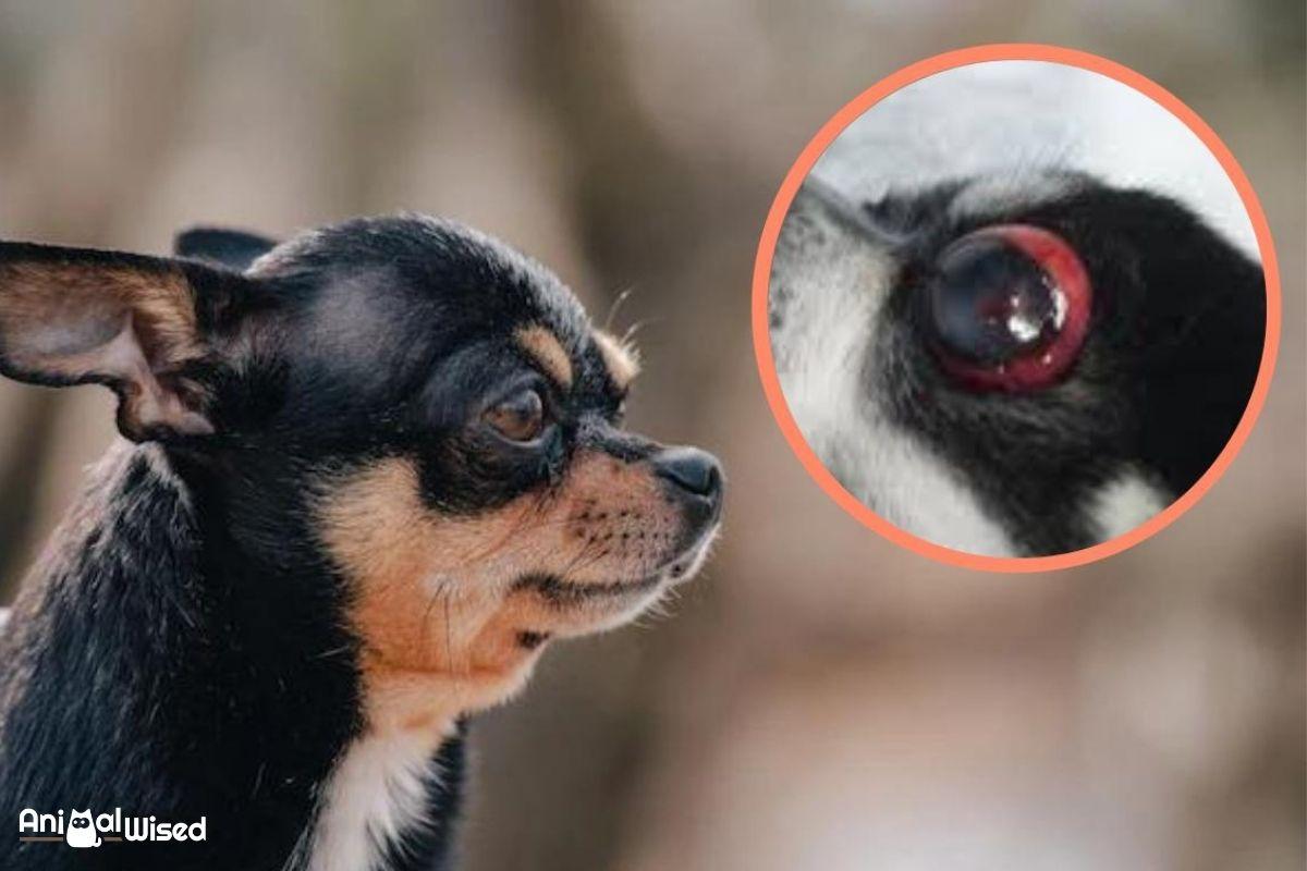
Ocular proptosis is one of the most important ophthalmological emergencies in the canine species. It consists of the forward displacement of the eyeball. This most commonly occurs as a result of trauma that causes the eye to exit its orbit, i.e. it pops out of its socket. Although it can appear in any breed, it is especially common in brachycephalic dogs.
If you see that my dog's eye popped out of socket, it can be traumatizing for us a dog guardians. This is why AnimalWised explains more about ocular proptosis in dogs, specifically is causes, treatment and recovery information. We will also take about symptoms and diagnosis of this ocular condition.
What is ocular proptosis in dogs?
Otherwise known as exophthalmos or exophthalmus, ocular proptosis occurs when the eyeball exits from its orbit, i.e. the socket in the skull which contains the eye and its appendages. The eye pops out of socket due to some type of force, most commonly trauma. Immediately after the eyeball moves from its normal position in the orbital space, the eyelids close over the area. In doing so, they prevent the eyeball from returning to its original position.
As a consequence of the eyeball's anterior movement, the following occurs:
- Venous return is impaired, causing conjunctival congestion.
- The cornea dries out and severe ulcers (exposure keratitis) develop.
- Associated uveitis occurs.
- Rupture of the extraocular muscles may occur and consequently a subconjunctival hemorrhage occurs.
- The optic nerve can be affected by the trauma itself or by the ensuing inflammation.
- Eventually, blindness will occur.
It is important to note that ocular proptosis is always an ophthalmologic emergency that requires immediate medical and surgical treatment. This reduces the risk of irreversible vision loss, i.e. blindness in dogs. Even in cases where vision cannot be recovered, carrying out prompt and correct treatment will allow the eyeball to be preserved and the aesthetics of the animal to be maintained. It also helps to prevent secondary infections and stress in the dog.

Causes of ocular proptosis in dogs
When a dog's eye has popped out of socket, the actual movement out of the orbital area will happen all at once. However, the process by which orbital anterior movement of the eyeball occurs may be progressive. This is due to various reasons. They include:
- Blows to the skull
- Traffic collisions
- Fights with other animals
- Falls
- Tumors
Most commonly, ocular proptosis is caused by trauma. Whether from a fight, traffic collision or any other type of trauma, the force of the impact will knock the eye from the socket. However, if a tumor develops behind the eye, the accumulation of cells can grow to the point it pushes the eyeball from the orbit.
Ocular proptosis most frequently occurs in brachycephalic dog breeds. These are dogs which have a flattened snout and shorter soft palate. Their flattened skull shape makes it more difficult for the eyeball to remain in its orbit, resulting in a bulging appearance of the eyes. When trauma occurs, it is easier for these breeds to develop ocular proptosis since the eye is already in a more vulnerable position.
When a dog has other diseases which affect the eyes, it is possible they can increase the risk of ocular proptosis. These include canine hydrocephalus which causes the eyes to bulge in their sockets. Leishmaniasis in dogs also has symptoms which affect the eye and cases of exophthalmos development have been seen[1].
Symptoms of Ocular Proptosis in Dogs
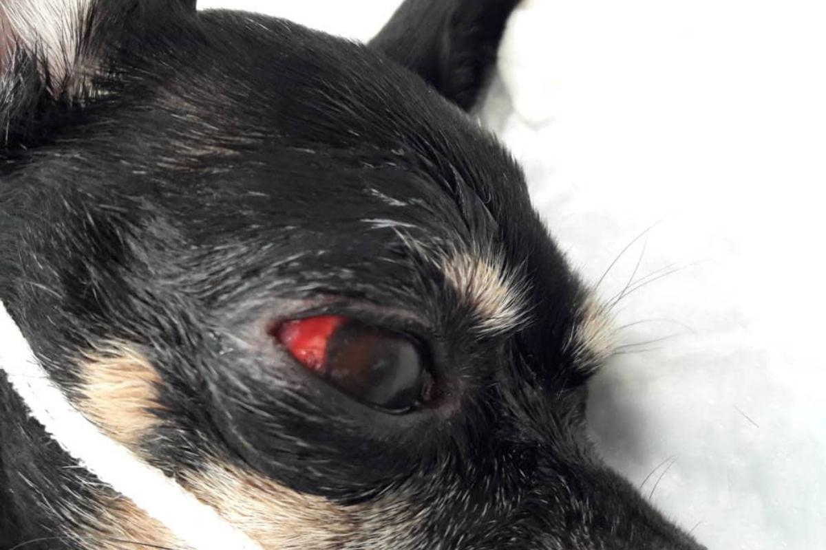
The most obvious sign of ocular proptosis in dogs is the fact that the eyeball has popped out of socket. However, we can also see the following symptoms appear with this condition:
- Edema and inflammation of the eyelids
- Desiccated cornea
- Corneal ulcers
- Chemosis (edema in the conjunctiva)
- Subconjunctival hemorrhage
- Hyphema (blood in the anterior chamber)
- Miosis (contraction of the pupil)
- Mydriasis (dilation of the pupil)
The pupil will either contract or dilate, not both at the same time. Hyphema can be seen with blood in the dog's actual eye, although you may see some blood around the exposed tissue.
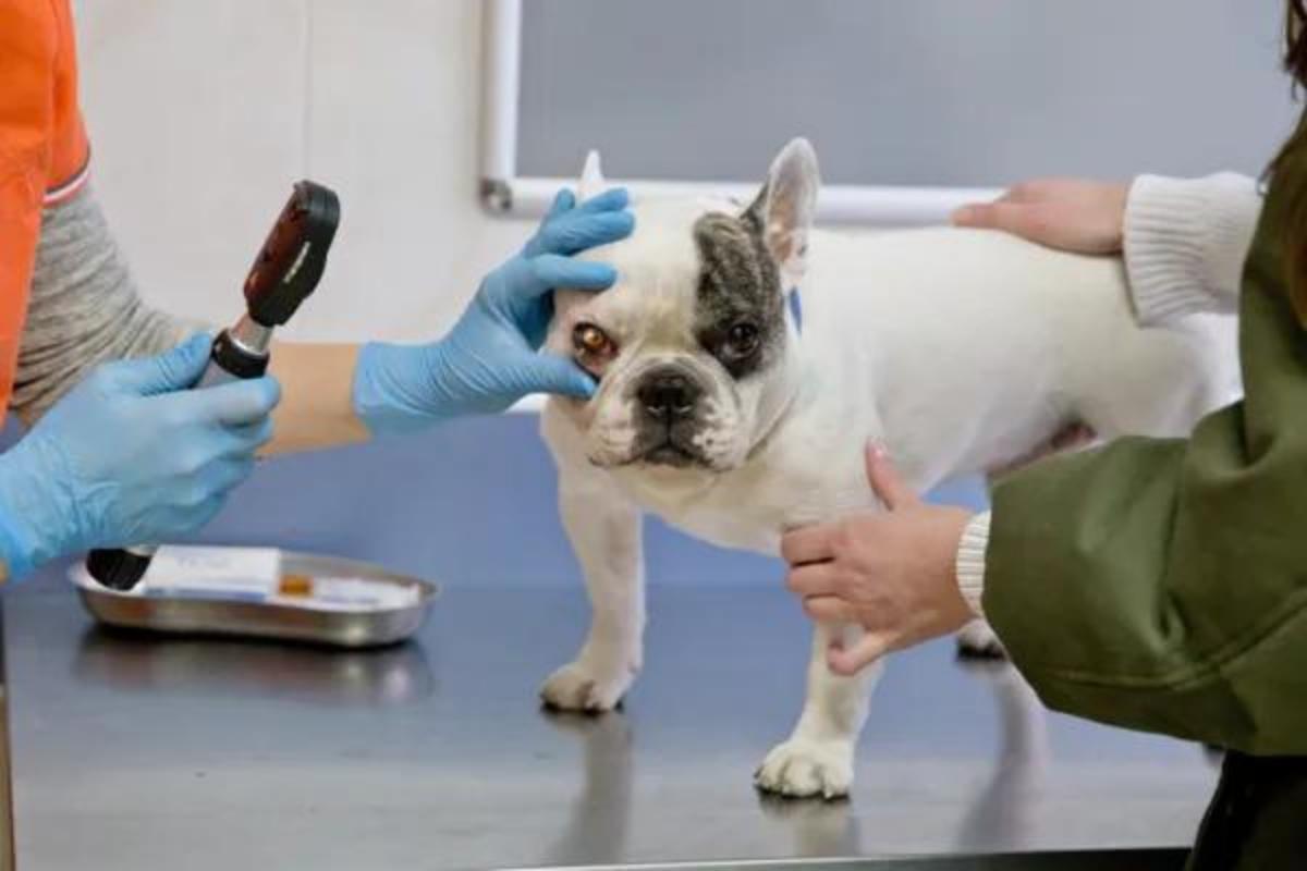
Diagnosis of ocular proptosis in dogs
Although ocular proptosis can be seen with physical examination due to the eye being popped out of socket, we will need to determine the extent of the damage causes. For this reason, the diagnosis of canine ocular proptosis should include the following points:
- Ophthalmological examination: a systematic examination of all the ocular structures must be carried out to assess the functionality of the eye in order to guide the treatment and issue a prognosis.
- Skull X-ray: when it occurs as a result of trauma, it is important to perform a head X-ray to rule out possible skull fractures or tumors. In addition, it is especially relevant to know if there is a fracture at the level of the orbit, since in this case it will not be possible to put the eyeball back in its place without the orbital structure healing.
- General examination: the general condition of the patient must be assessed and any systemic alteration produced by the trauma that could compromise the life of the animal should be ruled out.

Treatment of ocular proptosis in dogs
As we have stated in previous sections, ocular proptosis is an ophthalmological emergency that requires immediate treatment to avoid irreversible loss of vision or, enucleation (removal of the eyeball). It is important to act as quickly as possible.
Caregivers who detect or suspect possible ocular proptosis in their dog should administer first aid. This consists of protecting the eye from drying out and preventing further damage. Gauze should be moistened with serum or water and placed on the prolapsed eyeball to keep it protected.
You should then go to an emergency veterinary center. The veterinary team will be in charge of establishing a stabilizing treatment to prevent the process from worsening.
Medical therapy for ocular proptosis includes:
- Continuous irrigation of the cornea with saline solution to keep it moist and protection with cold compresses.
- Administer intravenous corticosteroids to reduce inflammation.
- Dehydrate the vitreous humor (clear fluid of the eyeball) to make the eyeball softer and easier to reintroduce into the orbital space.
- If the prolapse is partial, the animal can be sedated and the eyeball can be reintroduced manually. In no case should it be forced back in to the socket. In most cases, manual reintroduction does not usually work and it is necessary to resort to surgical treatment.
Surgical treatment of ocular proptosis in dogs involves repositioning the eyeball in the orbit under general anesthesia. Once reintroduced, the upper eyelid must be sutured to the lower one (known as tarsorrhaphy or blepharorrhaphy) to keep the eye closed for 15-20 days. If this is not performed, the eye would immediately prolapse again as a result of inflammation.
In some cases it is not possible to preserve the eyeball and it is necessary to perform an enucleation, i.e. removal of the eyeball. Generally, enucleation is recommended in the following cases:
- Rupture of more than 2 extraocular muscles
- Avulsion of the optic nerve (partial or complete section of the nerve)
- Corneal or scleral break
- Hyphema
- Severe sequelae 20 days after tarsorrhaphy
Ocular proptosis in dogs is a relatively rare eye condition. Learn about more common possibilities with our guide to common eye diseases in dogs.
Recovery from ocular proptosis in dogs
During the recovery or postoperative period, treatment should be instituted to combat inflammation and prevent infection:
- Apply cold compresses immediately after surgery.
- Administer systemic anti-inflammatory drugs (usually corticosteroids or NSAIDs ) for 7-10 days to reduce intraocular, eyelid and optic nerve inflammation. It is not recommended to administer them by ophthalmic route (in eye drops) since the cornea may be inflamed.
- Administer systemic and/or topical antibiotics.
Tarsorrhaphy or blepharorrhaphy should be maintained for between 15-20 days. After that time, the sutures must be removed and the possible sequelae in the affected eye must be evaluated.
Sequelae of ocular proptosis in dogs
Sequelae are pathological conditions resulting from disease or trauma. Canine ocular proptosis can occur, depending on the severity of the process. Specifically, the possible consequences in the affected eye that can occur include:
- Blindness
- Permanent squinting
- Corneal ulcers
- Dry keratoconjunctivitis
- Exposure keratitis
- Glaucoma
- Phthisis bulbi (atrophy of the eyeball)

Prognosis of Ocular Proptosis in Dogs
The prognosis for preservation of vision is guarded to severe. Most dogs with prolapsed eyes which have popped out of their socket experience irreversible blindness due to optic nerve damage or intraocular damage. In fact, only 20% of dislocated eyes retain their vision.
While every effort should be made to preserve vision, it should be noted that dogs can adapt to one-eyed vision without drastically affecting their quality of life. In rare cases of bilateral ocular proptosis (i.e. affecting both eyes), total blindness is likely.
This article is purely informative. AnimalWised does not have the authority to prescribe any veterinary treatment or create a diagnosis. We invite you to take your pet to the veterinarian if they are suffering from any condition or pain.
If you want to read similar articles to My Dog's Eye Popped Out of Socket, we recommend you visit our Eye problems category.
1. Peña, M. T., Roura, X., & Davidson, M. G. (2000). Ocular and periocular manifestations of leishmaniasis in dogs: 105 Cases (1993-1998). Veterinary Ophthalmology, 3(1), 35-41.
https://doi.org/10.1046/j.1463-5224.2000.00106.x
- Association of Spanish Veterinary Specialists in Small Animals. (AVEPA). Ophthalmological emergencies.
- Mejia, E. (2008). Orthopedics, neurology and rehabilitation in small species: dogs and cats. The Modern Handbook.


