Pleural Effusion in Cats - Causes, Symptoms and Treatment


See files for Cats
Pleural effusion is an accumulation of fluid in the pleural cavity, the space between the pleural sacs of each lung. There is some fluid naturally in this cavity, but when fluid builds up, it poses serious health risks. An abnormal buildup of pleural fluid can cause the cat respiratory distress. When this distress is sufficiently acute, it can cause the cat to hyperventilate and die. It is important to know that pleural effusion is not a disease in itself, but a symptom of other feline health disorders.
At AnimalWised, we look into more detail at pleural effusion in cats. We understand the causes, symptoms, and treatment of this fluid buildup so you can know both how to recognize it and what to do if it happens to your cat.
What is pleural effusion in cats?
Pleural effusion is an abnormal accumulation of fluid of various natures in the pleural cavity. Otherwise known as the pleural space, this is the area between the visceral pleura (the membrane that covers the lungs) and the parietal pleura (the one that covers the walls of the chest, mediastinum, and diaphragm). It naturally contains a minimal amount of fluid to lubricate the lungs during respiratory movements.
A disorder in the production or elimination of this fluid causes an excessive accumulation of it in the pleural space. This results in a restriction of lung movement during inspiration (lung expansion) that can cause the lobes of the lung to collapse. If left unchecked, this condition can lead to severe respiratory complications.
In general, pleural effusion in cats can be caused by any of the following mechanisms:
- Increased capillary permeability
- Reduction of the oncotic pressure of the capillaries
- Increased hydrostatic pressure in the capillaries
- Lymphatic obstruction.
To understand more about internal disorders which can lead to pleural effusion, take a look at our article on diaphragmatic hernias in cats. Moreover, recognizing the signs early can provide a better prognosis for affected cats.
Types of pleural effusion in cats
Pleural effusion in cats can be of various types depending on the nature of the fluid accumulated in the pleural space and the underlying cause of the effusion. To understand what type it is, the fluid must be analyzed. Once analyzed, it can be categorized according to a series of characteristics and parameters into the following:
- Pleural effusion of pure transudate: the color of the liquid is clear or yellowish, with a small amount of protein (<2.5 gr/dl), no fibrin, and little cellularity (<1,000 cells/microliter).
- Pleural effusion of modified transudate: with a somewhat cloudy yellowish-pink color, it has a protein quantity between 2.5 and 5 gr/dl, no fibrin, triglycerides, or bacteria, and a cell count of 1,000-15,000 cells/microliter (reaching to 100,000 if produced by lymphosarcoma). It also contains mesothelial cells, non-degenerated neutrophils, and neoplastic cells in lymphosarcoma.
- Pleural effusion of inflammatory exudate: it is the same color as the previous type, but the amount of protein is 2.5-6 gr/dl, even reaching 8.5 gr/dl in the case of feline infectious peritonitis (FIP). Fibrin is present, but without triglycerides or bacteria and a cell content of 1,000-20,000 cells/microliter (reaching 100,000 if produced by lymphosarcoma) and with non-degenerated neutrophils, macrophages, and neoplastic cells in tumors.
- Pleural effusion of septic exudate: a cloudy or opaque yellowish-brown color, the total proteins are 3-7 gr/dl and it contains fibrin and bacteria, but not triglycerides. The cell count is 5,000-300,000 cells/microliter and contains degenerated neutrophils, macrophages, and bacteria.
- Pleural lymph effusion: the color in this case is milky-white (although sometimes it can be pinkish-reddish) with a protein quantity of 2.5-6 gr/dl. It contains fibrin and triglycerides, but not bacteria. The cell content is 500-20,000 cells/microliter and usually contains lymphocytes, neutrophils, and macrophages.
- Pleural blood effusion: the color is red and opaque. It has more than 3 gr/dl of protein and fibrin, but not triglycerides or bacteria. The cell count is similar to that of peripheral blood and with red blood cells and some bank cells.
Proper identification of the type of pleural effusion is crucial for determining the appropriate treatment and managing the cat's condition effectively.
Causes of pleural effusion in cats
There are many causes that can produce an accumulation of fluid in the pleural space of cats. In general, any of the following diseases and disorders can cause feline pleural effusion:
- Liver disease: due to the development of hypoproteinemia that reduces oncotic pressure and allows fluid to leak out and accumulate in the pleural space.
- Kidney disease (glomerulonephritis): due to loss of protein in the urine. Discover here 4 symptoms of kidney disease in cats.
- Enteropathy: due to loss of proteins by the intestines.
- Congestive cardiomyopathy: due to congestive heart failure in diseases in cats such as congenital heart defects, feline dirofilariosis, hypertrophic cardiomyopathy, or pericardial diseases.
- Wet feline infectious peritonitis (FIP): due to an immune vasculitis, causing damage to the endothelium of the blood vessels and egress of proteins and serum from the capillaries. The exudate is non-septic fibrinous (nonbacterial).
- Bacterial infections: can cause the accumulation of pus (pyothorax) due to the entry of bacteria through bites and wounds, perforation of the esophagus or trachea, extension of pneumonia, penetration of foreign bodies, serious periodontal infection, etc.
- Tumors in the mediastinum: such as lymphosarcoma, thymoma, hemangiosarcoma, or breast tumors in cats.
- Lung tumor (adenocarcinoma): either primary or secondary due to metastasis from another location.
- Diaphragmatic hernia: usually due to trauma.
- Lung torsion: of the right or left middle pulmonary lobe.
- Chest trauma: due to lung injury or rupture of blood vessels in the chest, it produces pleural blood effusion (hemothorax), as well as rodenticide poisoning (coagulopathy).
In addition to these causes, environmental factors such as exposure to toxins and pollutants can exacerbate the risk of developing pleural effusion, especially in urban areas.
Symptoms of pleural effusion in cats
The main symptom of pleural effusion is the presence of fluid in the pleural cavity. However, since this is an internal process, we need to look at other clinical signs which can indicate the presence of this fluid:
- Dyspnea (respiratory distress)
- Reduced lung sounds
- Tachypnea increased (respiratory rate)
- Coughing
- Exercise intolerance
- Anorexia and weight loss
Depending on the disease or condition that caused the pleural effusion, the cat will present symptoms associated with the underlying process. For example:
- Congestive heart failure: cats will also have hypothermia, weak pulse, and jugular vein distention, as well as an enlarged liver and ascites.
- Feline infectious peritonitis: they may present depression, fever, and jaundice (yellowing of the mucous membranes), neurological and ocular signs.
- Mediastinal tumors: regurgitation and dysphagia may also appear due to compression of the esophagus, feline Horner's syndrome if the sympathetic chain of nerve fibers is compressed, edema of the neck and head if the cranial vena cava is compressed, reduction of heart and lung sounds, and distension of the jugular vein.
- Glomerulonephritis: cats will show signs of kidney disease such as increased urination and water intake, pale mucous membranes, vomiting, or uremic syndrome, among others.
- Liver disease: jaundice, increased liver enzymes, and ascites can be seen. In protein-losing enteropathy, edema and ascites can also be seen, as well as thromboembolic disease due to the loss of antithrombin at the intestinal level.
Additionally, some cats may exhibit lethargy and a noticeable change in behavior, such as hiding or avoiding interaction, which can signal discomfort or distress associated with pleural effusion.
Diagnosis of pleural effusion in cats
The cat's guardian will need to take the affected feline to the veterinarian and relay as much about their medical history as possible. A physical examination of the cat will first be carried out to note clinical signs, body condition, respiration, auscultation, and mental status.
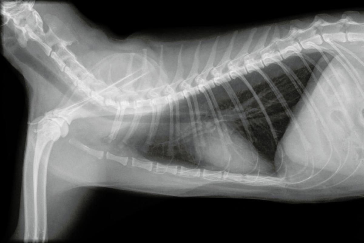
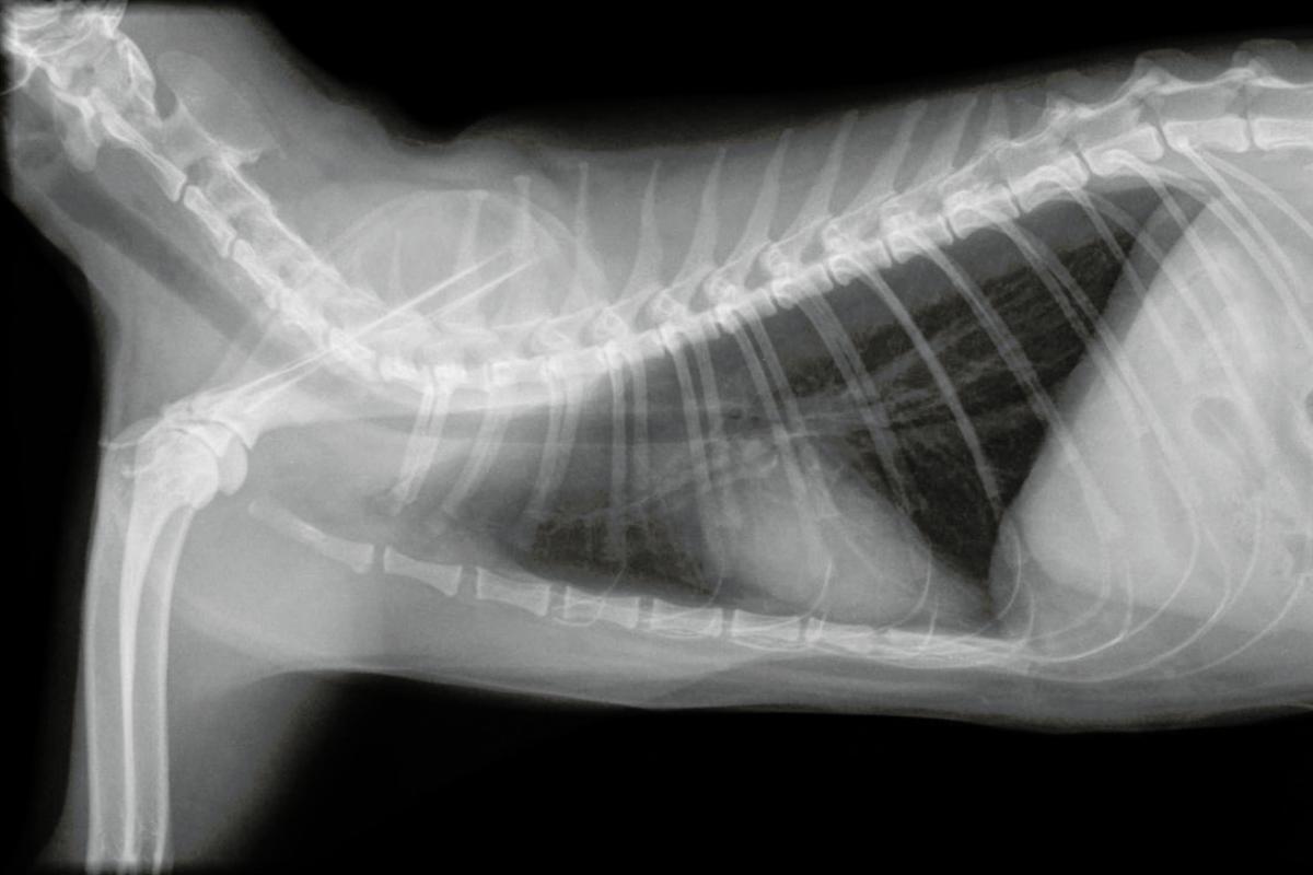
With symptoms such as respiratory distress, tachypnea, and reduced lung sounds, the diagnosis of pleural effusion is very likely. In an x-ray, the presence of fluid in the pleural space can be seen by preventing a normal visualization of the lungs. It is possible to suspect or deduce what type of fluid it is (transudate, blood, lymph, pus) with an ultrasound. However, analysis of fluid after thoracentesis by cell count, cytology, and biochemistry will be required to determine the type of pleural effusion. In case of suspected infection, the fluid should be cultured.
Other ways to diagnose pleural effusion in cats are:
- Electrocardiogram: to assess heart function and detect arrhythmias and tests for FIP virus in suspected cases of this infectious disease. Advanced diagnostic tools like CT scans can also provide detailed images of the chest cavity, aiding in a comprehensive assessment.
- Blood tests, biochemistry, and urinalysis: they are essential to rule out kidney, liver, or digestive causes and observe the general state of health of the cat.
Early diagnosis and intervention are critical in managing pleural effusion effectively, minimizing the risk of severe complications.
Treatment of pleural effusion in cats
Pleural effusion therapy will depend on its underlying cause. Emergency treatment includes oxygen therapy due to respiratory distress, puncture of the pleural space to drain the fluid (thoracentesis), samples to be taken for analysis, and diuretics such as furosemide or spironolactone to reduce the stress of the cat. This treatment will depend on the origin of the cause.
- In tumors: chemotherapy should be used. In some tumors, as well as diaphragmatic hernias and torsion of the pulmonary lobe, the treatment will be surgical.
- In case of pyothorax: the infection that is causing the accumulation of pus in the pleural space should be treated with antibiotics. In chylothorax due to accumulation of lymph in the pleural space, the chyle should be drained frequently by thoracocentesis or by placing a drainage tube in the cat. If this is not effective, surgical treatment should be considered with ligation of the thoracic duct after draining the lymph from the pleural cavity.
If heart failure is evident, in addition to diuretics and oxygen, drugs such as nitroglycerin or digoxin can be used. In renal, hepatic, and intestinal disease, an effective therapy must be adapted to control said pathologies.
Moreover, supportive care, including nutritional support and fluid therapy, can enhance recovery and improve the overall prognosis for cats suffering from pleural effusion. Regular monitoring and follow-up visits are crucial to ensure the effectiveness of the treatment plan.
Sequelae of a pleural effusion in cats
Pleural effusion in cats can leave sequelae (conditions ensuing from the underlying cause). In general, with proper therapy and diagnosis of the problem, cats maintain their health and quality of life as before the effusion. Among the main sequelae of pleural effusion in cats, we find:
- Lung damage: such as pulmonary edema, which can affect respiratory efficiency.
- Poorly resolved infection: develops into a chronic abscess called an empyema, requiring further intervention.
- Presence of air in the chest cavity: pneumothorax after thoracentesis, which may necessitate additional medical procedures to resolve.
It should be noted that vigilant post-treatment care and regular veterinary check-ups play a vital role in preventing long-term complications and ensuring a good quality of life for affected cats.
This article is purely informative. AnimalWised does not have the authority to prescribe any veterinary treatment or create a diagnosis. We invite you to take your pet to the veterinarian if they are suffering from any condition or pain.
If you want to read similar articles to Pleural Effusion in Cats - Causes, Symptoms and Treatment, we recommend you visit our Breathing diseases category.
- Aybar, V., Casamian, D., Ceron, J. J., Clement, F., Fatjó, J., Lloret, A., Lujan, A., Novellas, R., Perez, D., Silva, S., Smith, K., Tegles, F., Vega, J., & Zanna, G. (2018). Clinical Manual of Feline Medicine. Ed.SM Publishing LTD. Sheffield, UK.