Sialocele in Dogs - Causes, Symptoms and Treatment


See files for Dogs
Certain dogs are known for the considerable amount of drool they produce. While this provides practical considerations in terms of home hygiene, we may not think too much about where the drool comes from. A dog's salivary glands are very important, even if some are put to work more than others. Although diseases of the salivary glands are rare in small mammals, a sialocele in dogs is a relatively common pathology in dogs. Also known as a salivary mucocele in dogs, it is an accumulation of saliva which forms a cyst in and around the dog's mouth.
At AnimalWised, we find out more about the causes, symptoms and treatment of sialocele in dogs. We find out how you can detect salivary mucoceles and what sort of treatment options you might expect.
What is a sialocele in dogs?
Sialocele is an accumulation of saliva in subcutaneous tissue due to damage of the salivary gland or its ducts. It is commonly associated with physical trauma to glands, but it is usually a benign condition. It is also known as a salivary mucocele. The dog's saliva collects in the subcutaneous tissue, resulting in an inflammatory reaction. This causes the sialocele to be lined by granulation tissue (inflammatory tissue).
Although all breeds of dog are susceptible to this pathology, sialoceles are more common in certain breeds. These breeds include the following:
- German Shepherd
- Dachshund
- Miniature Poodle
- Australian Silky Terrier
They mainly occur in animals under 4 years of age. There are different types of salivary mucocele in dogs, with cervical sialocele being the most common. This type of canine sialocele has a global incidence rate estimated to be around 0.5%. Furthermore, some studies suggest that environmental factors and genetic predispositions may also influence the likelihood of developing sialocele, but more research is needed to confirm these theories.
Types of sialocele in dogs
As stated above, there are different types of salivary mucocele in dogs. The type is mainly determined by location. Dogs have four main pairs of salivary glands. These are parotid, mandibular, sublingual and zygomatic. When the sialocele originate in different glands, they are classified into the following:
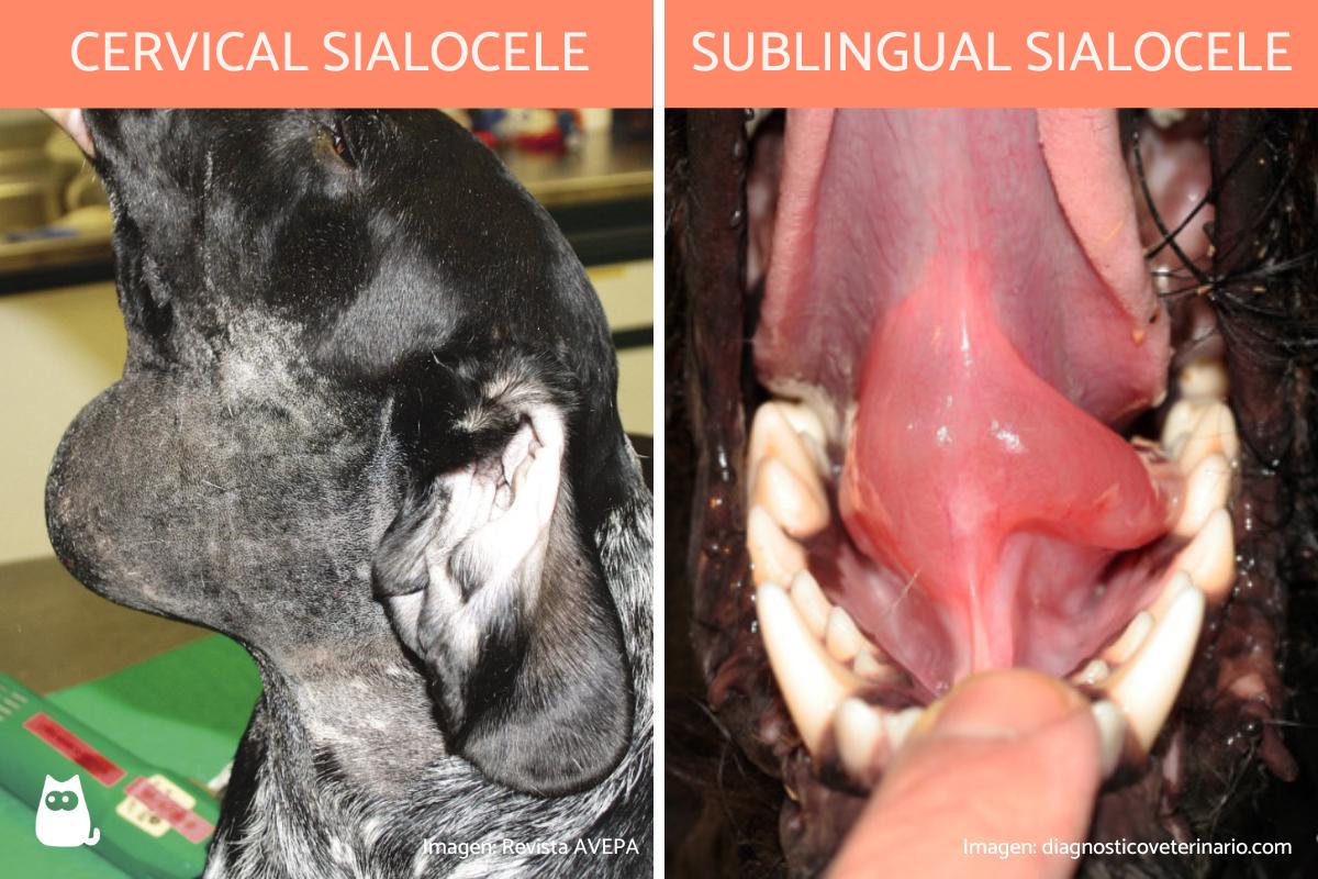
- Cervical sialocele: appears in the intermandibular space or in the upper area of the neck. It occurs when the mandibular and/or sublingual salivary glands are affected. It is the most common presentation of salivary mucocele in dogs[1].
- Sublingual sialocele: commonly known as a ranula in dogs. In this case, the salivary mucocele forms on the floor of the mouth, to one side of the tongue.
- Pharyngeal sialocele: a variation of the cervical mucocele, in which saliva accumulates mainly at the level of the pharynx. This type of sialocele is much less common.
- Zygomatic sialocele: one that originates in the zygomatic salivary glands, which are located just below the eye. This is a very rare mucocele.
In addition to these, veterinarians have observed cases where sialoceles develop in atypical locations, further complicating the diagnostic process. Such occurrences underscore the importance of professional veterinary consultation.
What is the difference between a sialocele and a mucocele in dogs?
To a certain extent, we can use the terms ‘sialocele’ and ‘salivary mucocele’ interchangeably. However, it is important to note that an oral mucocele refers to various types of polyp which can affect a dog's mouth. They are caused by damage to the salivary glands. A sialocele is a variant of a sialocele that develop from the leakage of saliva from the gland. In this way a sialocele is a salivary mucocele, but not all oral mucocele are sialoceles.

Symptoms of sialocele in dogs
Salivary mucocele in dogs is associated with the appearance of a soft, fluctuating, painless, and cold mass (like a soft cyst). Recognizing these four characteristics will be very important to diagnose the process. It is using these criteria that a veterinarian will be able to differentiate between a sialocele and an inflammation of the salivary glands in dogs.
Generally, the mass associated with the sialocele grows slowly and does not cause other associated problems. However, there is the possibility of certain complications:
- When the salivary mucocele is sublingual, dogs may have difficulty chewing and swallowing food. They may even bleed if there is trauma to the mucocele while chewing.
- When the salivary mucocele is pharyngeal, difficulty swallowing (dysphagia) or even respiratory distress may appear. This is due to compression or obstruction of the airways caused by the inflammation.
In rare cases, the presence of a sialocele can lead to secondary infections if the cyst ruptures and becomes contaminated. This makes it crucial for pet owners to act swiftly if they notice any suspicious swelling.
Learn about another type of swelling around the dog's mouth and neck with our article on whether dogs can get mumps.
Causes of sialocele in dogs
In most cases, sialoceles in dogs occur as a result of trauma that causes a ruptured salivary gland or duct. These injuries are usually associated with pulling too hard on the leash, fights between dogs, falls and chewing on hard or sharp objects, among others.
Although some are less common than others, there are also other possible causes of sialocele in dogs. These include:
- Surgical complication: when the dog's salivary gland has been operated on, trauma from the surgical intervention can damage the gland and result in a sialocele. Other oral surgeries may also cause damage to the salivary gland.
- Tooth extraction: trauma from the extraction can damage the salivary gland.
- Sialolithiasis: the formation of salivary stones (stones) can cause a duct or salivary gland to rupture in dogs with a sialocele as consequence.
- Foreign bodies: if a dog chews on a foreign object, splinters or other traumatic injuries can occur to the salivary glands.
- Neoplasms: benign or malignant tumors can grow on or around the salivary gland and result in sialocele.
- Idiopathic cause: a cause of unknown origin.
Additionally, some veterinarians have hypothesized that certain autoimmune conditions may play a role in the development of sialoceles, although this is not yet fully understood. Learn more about how neoplasms can affect canines with our guide to different types of tumors in dogs.
Diagnosis of mucocele in dogs
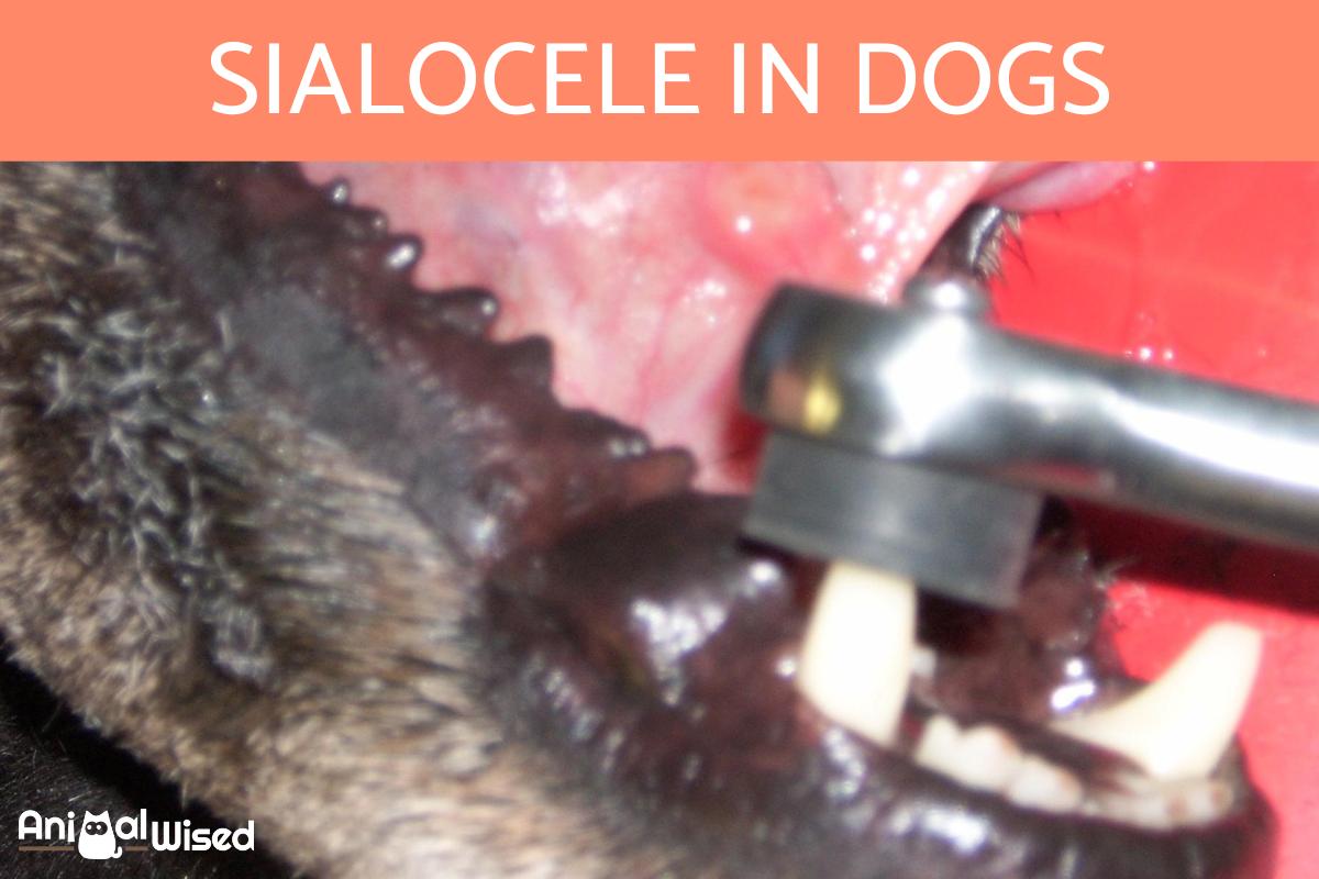
The diagnosis of salivary mucocele in dogs is based on examination of the symptoms. This is requires the detection of a soft, fluctuating, cold and painless mass in the area of projection of one of the dog's salivary glands. In the specific case of pharyngeal salivary mucoceles, it is necessary to perform an exploration of the oral and pharyngeal cavity under sedation.
To confirm the diagnosis, it is necessary to perform a puncture-aspiration of the gland. This is the use of a surgical needle which is inserted into the sialocele to release fluid. If it is a sialocele, a yellow or slightly blood-tinged, viscous, mucoid, transparent fluid with low cellularity will be obtained.
Other diagnostic techniques can be used to differentiate sialocele from other possible differential diagnoses such as tumors or cysts. These can include x-ray, ultrasound or biopsy. These will only be required if there is doubt after examination or aspiration. One recent study suggests that computerized tomography (CT) scan is useful as an adjunct diagnostic test for treatment planning of sialoceles[2]. Moreover, advances in imaging technology have enabled more accurate and less invasive diagnostic procedures, which can be particularly beneficial in complex cases.
Treatment of sialocele in dogs
Now we know more about the causes and symptoms of sialocele in dogs, we can look at what treatment options are available. Although it is a self-limiting complication which should not cause serious threat to the dog, it is not a problem that will resolve on its own.
The most common treatment of sialocele in dogs is a gradual process. Not only does aspiration with a surgical needle help us to diagnose the salivary mucocele, but it is used to drain the fluid. This is performed periodically, allowing the affected glands to close and heal over time. This type of treatment should take around 1 week for the salivary glands to heal.
When the aspiration does not work, it will be necessary to carry out surgical intervention. This is the most effective type of treatment of sialocele in dogs, but it is also invasive and will usually only be carried out when necessary.
Sialocele or salivary mucocele surgery in dogs
When periodic aspiration of the sialocele is not effective, surgical removal of the affected salivary glands will be required. This is a technique known as sialoadenectomy. The surgical treatment of this pathology has a favorable prognosis in almost 100% of the cases. Only 0.5% of the operated dogs present recurrences. These are usually associated with an incomplete or mistaken resection of the gland.
In the case of sublingual mucoceles, removal of the glands is usually combined with marsupialization. This is a process which consists of leaving a permanent opening in the lesion to facilitate its continuous drainage into the oral cavity.
Furthermore, post-operative care is crucial to ensure the success of the surgery and prevent complications. Regular follow-up visits to the veterinarian can help monitor the healing process and catch any signs of infection early.
If you observe any type of lump in the mouth, neck or similar area of your dog, it is vital you go to a veterinarian. They will be able to diagnose the condition and distinguish it from other types of lumps in a dog's neck. They will then administer the correct form of treatment. The cost of sialocele surgery (sialoadenectomy) in dogs will depend on the clinic, but it can range anywhere between $250 to $1000.
Home treatment of sialocele in dogs
Since it is a relatively benign condition, some dog guardians may wonder if they can treat salivary mucocele on their own at home. Treatment of sialocele must only be carried out by a professional veterinarian. You should not drain the fluid yourself as it needs to be carried out in sterile conditions with the proper clinical tools. If we do it at home, we risk a serious salivary gland infection which can be very painful, distressing and can even lead to blood poisoning.
Surgical removal of the sialocele can also not be carried out at home. This is not only due to hygiene issues, but also because the dog will need to be properly sedated and provided with appropriate follow-up treatment. If your dog has a sialocele, take them to a veterinarian.
It's important to remember that while home remedies can be tempting, they are not a substitute for professional veterinary care. Attempting to treat a sialocele at home could lead to complications that are far more serious than the initial condition.
This article is purely informative. AnimalWised does not have the authority to prescribe any veterinary treatment or create a diagnosis. We invite you to take your pet to the veterinarian if they are suffering from any condition or pain.
If you want to read similar articles to Sialocele in Dogs - Causes, Symptoms and Treatment, we recommend you visit our Other health problems category.
1. Landy, S., Peralta, S., & Fiani, N. (2021). An Atypical Presentation of a Zygomatic Sialocele in a dog. Journal of veterinary dentistry, 38(4), 223–230.
https://doi.org/10.1177/08987564211072675
2. Tan, Y. L., Marques, A., Schwarz, T., Mitchell, J., & Liuti, T. (2022). Clinical and CT sialography findings in 22 dogs with surgically confirmed sialoceles. Veterinary radiology & ultrasound: the official journal of the American College of Veterinary Radiology and the International Veterinary Radiology Association, 63(6), 699–710.
https://doi.org/10.1111/vru.13104
- Amaya, J., Avila, J. C. (2014). Cervical mucocele in a canine. Rev Zooc, 1(2), 3-6.
- Lebrero, M. E., Ramon, E., Alaman, M., Unzueta, A., & González, A. (2016). Cervical salivary mucocele caused by a sialolith in a dog. Clin Vet Peq Anim, 36(3), 179-183.


