Autoimmune Skin Disease in Cats - Pemphigus in Cats


See files for Cats
Although rare, cats can suffer from autoimmune skin diseases which includes pemphigus foliaceus, pemphigus vulgaris, pemphigus erythematosus and paraneoplastic pemphigus.
In this AnimalWised article we're going to talk about autoimmune skin disease in cats, including the different types of pemphigus in cats. Keep reading to learn more.
What is feline pemphigus?
Feline pemphigus is an autoimmune disease in which a cat's immune system does not recognize part of its body as its own and creates an immune reaction attacking it. It consists of cutaneous or mucocutaneous disorders due to a type II hypersensitivity reaction that begins with the participation of immunoglobulins G and M, which bind to target cells and activate complement, inducing phagocytosis. This leads to the production of autoantibodies against certain components of the epidermis.
Pemphigus in cats is a dermatological disease characterized by an acantholysis or detachment of each cell of the epidermis that creates vesicles within it. These vesicles can be infiltrated with eosinophils or neutrophils and become pustules.

Types of pemphigus in cats
There are different types of pemphigus in cats. They vary according to the distribution of the lesions and their pathological characteristics. The classifications are the following:
- Pemphigus vulgaris: consists of the formation of vesicles or blisters in the oral cavity, skin and mucocutaneous junctions, such as in the armpit and the inguinal region. These lesions, due to their fragility, evolve into collarettes, erosions, ulcers and scabs.
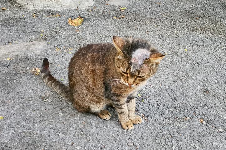
- Pemphigus foliaceus: autoantibodies are produced against the proteins of the stratum spinosum of the epidermis. This type is characterized by the formation of vesicles, bullae or, less often, subcorneal pustules that affect follicles and interfollicular skin. Secondary lesions are erythema, exudation, scabs, alopecia, and collarettes. These are generally distributed symmetrically on the face, muzzle, ears up to the extremities and the abdomen. The lesions occur on the skin, without affecting the oral cavity or mucocutaneous junctions.
- Pemphigus erythematosus: it is considered an intermediate form between pemphigus erythematosus and pemphigus foliaceus. Vesicles and blisters and pustular lesions form on the ears and head of the cat. It is to be considered that solar radiation can aggravate the pathology.
- Paraneoplastic pemphigus: this is when vesicles and blisters occur in multiple organs other than the skin. Paraneoplastic pemphigus is a disease associated with an underlying cancer, generally of lymphoproliferative origin.
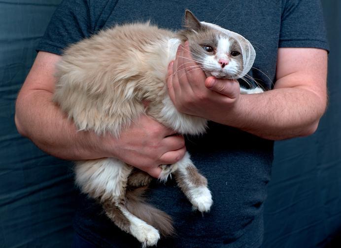
Symptoms of pemphigus in cats
Cats with pemphigus present, in addition to the lesions described above according to the type they develop, nonspecific signs such as:
- Fever
- Anorexia
- Lethargy
- Apathy
- Pain
- Lymphadenopathy
In cats, pemphigus foliaceus is the most common autoimmune disease . More than subcorneal pustules, which are more frequent in dogs, yellowish crusts are more frequently observed in cats with pemphigus foliaceus. A characteristic lesion of this pemphigus in cats is paronychia (inflammation of the skin around the nail) and pruritus (itching).

Diagnosis of feline pemphigus
Due to the itching produced by pemphigus foliaceus in cats, a differential diagnosis must be made between other diseases that cause itching in this species, such as allergies and parasitic diseases. In addition, the following tests will be carried out:
- Blood test : when faced with this type of injury in a cat, a blood test should be started, which may be normal or show an increased neutrophil and eosinophil count. Blood chemistry is normal if there is no concurrent disease.
- Cytology : Cytology of the lesions can aid in the diagnosis if neutrophils and acantocytes are seen. It is also useful to assess whether there is a bacterial infection. In that case, the cat would be treated with antibiotics before taking and sending the biopsy to the laboratory.
- Histopathological examination : however, the definitive diagnosis is achieved with histopathological examination. To do this, biopsies of recent primary lesions must be collected, being important that the cat has not received immunomodulatory or immunosuppressive treatment in the previous days, as it may alter the results. The biopsy will find subcorneal pustules with neutrophils and a variable number of acantocytes and eosinophils. If these are not observed, a presumptive diagnosis can be made if serocellular scabs are seen with acantocytes and neutrophils.
As a curiosity, in 90% of diagnoses of pemphigus vulgaris, oral lesions are detected. Paronychias would be seen in 30% of pemphigus foliaceus and itching in 80%.
Treatment of pemphigus in cats
Treatment should contain immunosuppressive drugs such as prednisolone at a dose of 2-8 mg / kg every 24 hours orally. Immunosuppressive doses should be reduced when remission of clinical signs begins, to the lowest dose that will keep the disease in resolution.
If the clinical signs are not reduced one month after the start of immunosuppressive treatment, it is recommended to switch to dexamethadone or methylprednisolone , decreasing to the minimum effective dose.
If no response is observed with these treatments or side effects such as polyphagia, polyuria-polydipsia, apathy, diarrhea, diabetes or urinary tract infections appear, chlorambucil should be added (0.1-0.2 mg / kg every 24-48 h). In some cases, corticosteroids can be suppressed and maintained with chlorambucil alone twice a week or every other day. The beneficial effects of this drug can take weeks to appear. It should be taken into account that chlorambucil is a cytotoxic drug, so periodic blood tests should be performed every 2-4 weeks for the first 3 months, until passing to every 6 months thereafter.
On the other hand, it has been determined that the use of cyclosporine at doses of 4.4 to 7.4 mg / kg every 24 hours can be effective for feline pemphigus, even being able to suppress corticosteroids and with efficacy similar to chlorambucil.
Additionally, drugs for cats with pemphigus may also contain immunomodulators such as mycophenolic acid and leflunomide.

This article is purely informative. AnimalWised does not have the authority to prescribe any veterinary treatment or create a diagnosis. We invite you to take your pet to the veterinarian if they are suffering from any condition or pain.
If you want to read similar articles to Autoimmune Skin Disease in Cats - Pemphigus in Cats, we recommend you visit our Skin problems category.
- G. Machicote. (2011). Canine and feline dermatology. Servet.
- Avepa. (2016). Day-to-day dermatology. Available at: https://www.avepa.org/pdf/proceedings/DERMATOLOGIA_2016.pdf


