Histiocytoma in Dogs - Causes, Symptoms and Treatment


See files for Dogs
Cutaneous histiocytoma is a tumor that frequently occurs in young dogs less than 4 years of age. It is a tumor that appears in the form of reddish hairless nodules, generally in the head and neck area. Once they appear, the lesions grow rapidly, often causing great concern for caregivers. Despite their threatening appearance, it is important to know that it is a benign tumor. It does not have aggressive behavior and most commonly resolves itself spontaneously after a few weeks.
If you want to know more about histiocytoma in dogs, AnimalWised explains its causes, symptoms and treatment in more detail. You can also see pictures of the condition throughout the article.
What is a histiocytoma in dogs?
Histiocytoma is a tumor that originates from histiocytes, a group of lymphoid cells that are an essential part of the dog's immune system. Cutaneous histiocytoma is a very common benign tumor in dogs. One report from the United Kingdom claims that histiocytoma is the most common single tumor type in dogs[1]. Although it can occur in older dogs, the prevalence of this type of dog tumor drops significantly after 3 years of age[2].
Certain dog breeds are more prone to developing histiocytoma than others. Although it can develop in any purebred or mixed-breed dog, the following dog breeds are most likely to develop histiocytoma:
- Boxer
- Boston Terrier
- Dachshund
- Bulldog
- Doberman
- Pug
Canine histiocytoma is a rapidly growing type of tumor which can become relatively large in only a few days. Fortunately, these types of tumors are benign neoplasms. They do not have aggressive behavior and resolve themselves spontaneously on their own.
When a guardian observes any sort of tumor on a dog's skin, it is very important to avoid misdiagnosis. With cutaneous histiocytoma, it is essential we do not confuse it with a fibrous histiocytoma in dogs. This is because fibrous histiocytoma can be malignant. It originates in the fibroblasts or undifferentiated mesenchymal cells. This is one of the reasons why we require veterinary diagnosis of any type of lump on a dog's skin.
In addition, understanding the typical behavior and characteristics of histiocytoma can help owners remain calm when they notice these growths, as they often resolve without intervention. Regular veterinary check-ups can further ensure any changes are monitored effectively.
Symptoms of histiocytoma in dogs
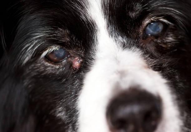
Cutaneous histiocytoma in dogs usually presents as a single nodule (up to 4 cm in diameter). Its appearance is usually pink or reddish in color and alopecic (i.e. hairless). An example of this type of tumor can be seen in the histiocytoma in dogs picture below. The appearance of multiple nodules is rare, although it has been described with some frequency in Shar Pei dogs.
Lesions associated with histiocytoma usually occur on the head (mainly in the ear pinna) and neck, although they can also appear on the extremities. They less frequently appear on the dog's trunk. Take a look at our related article to discover other reasons a dog has a lump on their neck.
Generally, these primary lesions do not cause discomfort in the dog. Sensations are usually limited to a slight itching. When the animal licks, scratches or rubs the nodule, it can ulcerate and become infected, causing mild local complications. Such complications include a lick granuloma in dogs.
Furthermore, the irritation from constant licking or scratching can exacerbate the nodule's size and slow down the natural healing process. In some cases, pet owners might notice a slight swelling in the area, which can be managed with proper veterinary care. Recognizing these symptoms early and consulting with a veterinarian can prevent further complications.

Causes of histiocytoma in dogs
In general, tumors arise as a consequence of a genetic mutation in the genome of a cell, triggering its exacerbated growth in the process. In some tumors the factor that triggers the genetic mutation in question is known (radiation, toxic compounds, hormones, etc.). In the majority of tumors this information is idiopathic, i.e. the tumors are of unknown origin.
This is the case of canine histiocytoma. Currently there is no information on what causes it, so it is considered an idiopathic tumor (i.e. of unknown origin).
Discover more about another common tumor in dogs with our article on the causes of canine mast cell tumors.
It should be noted that while the precise cause remains unidentified, ongoing research is exploring potential hereditary factors. Some studies suggest that certain breeds may have genetic predispositions making them more susceptible to developing these tumors. This information could eventually lead to targeted prevention strategies in the future.
Diagnosis of histiocytoma in dogs
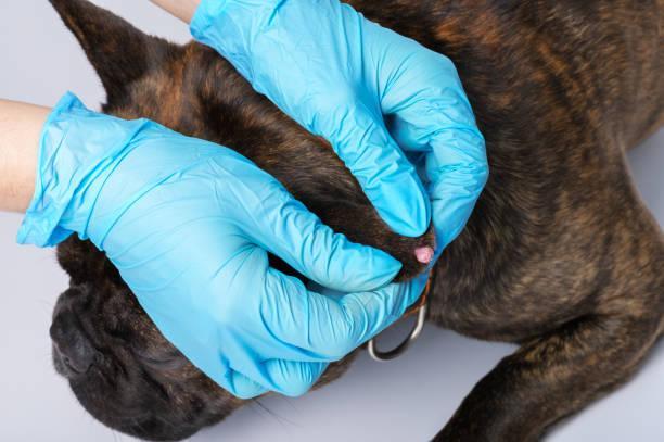
The appearance of rapidly growing, hairless, reddish nodular lesions are usually quite indicative of cutaneous histiocytoma. This especially when they appear on the head or neck of young dogs. To confirm the diagnosis it is necessary to perform a microscopic study of the lesion. To do this, a sample must be taken through fine needle puncture and cytology performed to confirm the cell line that caused the tumor.
In some cases, additional diagnostic imaging techniques such as ultrasound or MRI might be utilized to gain a better understanding of the tumor's structure and to rule out other potential conditions. These advanced diagnostic methods can provide a comprehensive view and assist in confirming the benign nature of the histiocytoma.
Treatment for histiocytoma in dogs
Cutaneous histiocytoma in dogs is a benign tumor that usually produces spontaneous regression in the weeks following its appearance. As a self-limiting neoplasm, we should know that there is no specific treatment. Cutaneous histiocytomas will progressively shrink in size and eventually disappear.
Despite the fact treatment is not usually required for histiocytoma, sometimes surgical removal is recommended. Your veterinarian may recommend excision of the histiocytoma tumor in the following circumstances:
- The location of the tumor is annoying for the animal (this usually happens when the tumor appears in the ear).
- The tumor takes longer than normal to disappear.
- Secondary complications appear (such as ulcers or bacterial infections) as a result of licking, scratching or rubbing the lesion.
- The cytological diagnosis is not clear and a more in-depth histopathological study is necessary.
In the event that secondary infections occur, it will be necessary to institute antibiotic treatment. Once the tumor has regressed or has been removed, the animal's prognosis will be favorable.
Additionally, maintaining good hygiene and monitoring the affected area can help prevent infection and promote faster healing. Owners should ensure their pets are comfortable and restrict activities that might irritate the nodule. In some cases, medications to reduce itching and inflammation may be prescribed to alleviate the dog's discomfort during the healing process.
Now that you know all the details about histiocytoma in dogs, you may be interested in learning about another type of benign tumor in dogs with our article on treating canine lipoma.
This article is purely informative. AnimalWised does not have the authority to prescribe any veterinary treatment or create a diagnosis. We invite you to take your pet to the veterinarian if they are suffering from any condition or pain.
If you want to read similar articles to Histiocytoma in Dogs - Causes, Symptoms and Treatment, we recommend you visit our Skin problems category.
1. Dobson, J. M., Samuel, S., Milstein, H., Rogers, K., & Wood, J. L. (2002). Canine neoplasia in the UK: estimates of incidence rates from a population of insured dogs. The Journal of small animal practice, 43(6), 240–246.
https://doi.org/10.1111/j.1748-5827.2002.tb00066.x
2. Moore, P. F., Schrenzel, M. D., Affolter, V. K., Olivry, T., & Naydan, D. (1996). Canine cutaneous histiocytoma is an epidermotropic Langerhans cell histiocytosis that expresses CD1 and specific beta 2-integrin molecules. The American journal of pathology, 148(5), 1699–1708.


