Melanoma in Dogs


See files for Dogs
Melanoma, a type of cancer developing from pigment cells, lurks in the shadows for many dogs. While not always aggressive, it can be a serious threat, particularly when left undetected. Knowing the early signs of melanoma in dogs is crucial, as prompt diagnosis and treatment significantly improve their chances of a positive outcome.
The following AnimalWised article explains what melanoma in dogs is, its symptoms, types, diagnosis, and options. By understanding this disease, you can become your dog's best health advocate and ensure they receive timely care for a better outcome.
What is melanoma in dogs?
Canine melanoma, a form of cancer, arises from melanocytes, specialized cells located in the skin's outer layer (epidermis) and within hair follicles. These melanocytes are responsible for producing melanin, the pigment that gives skin and hair their color. When these cells undergo abnormal growth and division, they form tumors known as melanomas.
These tumors can affect different layers of the skin. They may involve both the epidermis and dermis (the deeper layer of skin), or they may be confined to the dermis alone. In some cases, melanomas can grow to significant sizes, ranging from tiny growths measuring mere millimeters (about 0.04 inches) to sizable masses spanning up to 10 centimeters (approximately 3.94 inches) in diameter.
Regarding demographics, melanomas in dogs commonly appear in older animals, typically between the ages of 9 and 11 years. While there is no discernible gender predisposition, certain breeds exhibit a higher likelihood of developing these tumors than others.
Types of melanomas in dogs
There are various types of melanomas in dogs, categorized according to different factors including their location, behavior, and the presence of melanin. Let's examine each aspect more closely:
Location:
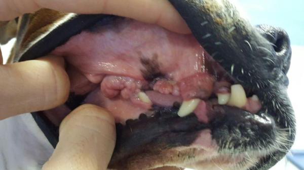
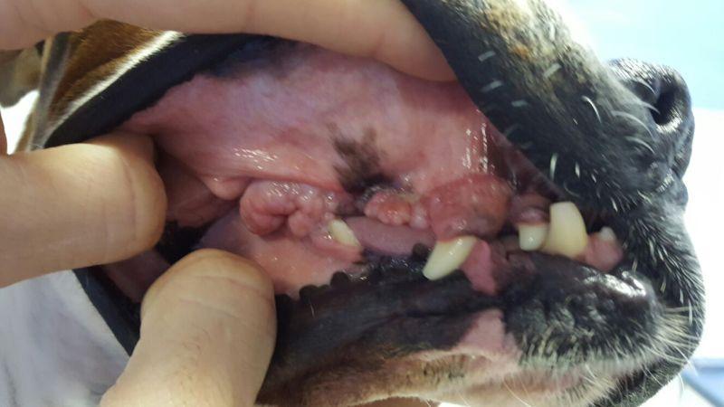
- Oral melanomas: this is the most prevalent type, constituting approximately 80% of cases. They manifest within the mouth, typically affecting the gums, tongue, lips, or palate.
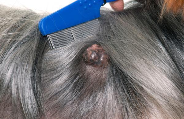
- Dermal melanomas: these occur on the skin anywhere on the body and may present as single or multiple lesions. Their appearance can vary from pigmented to non-pigmented.
- Nailbed Melanomas: developing on the nail bed or subungual crest (the area above the nail), these melanomas often produce symptoms such as limping, swelling, bleeding, or discharge from the affected toe.
- Ocular melanomas: less common, these melanomas affect structures within the eye. Symptoms may not be immediately noticeable, but they can lead to vision problems as they progress.
Behavior:
- Malignant melanomas: these are cancerous and have the potential to metastasize to other parts of the body, such as lymph nodes or lungs. The aggressiveness of malignant melanomas varies based on their location and specific subtype.
- Benign melanomas: these are non-cancerous and do not spread. However, they can still grow and cause discomfort, underscoring the importance of monitoring and potential removal.
Presence or absence of melanin:
- Pigmented Melanomas: these make up around 80% of cases and contain melanin, appearing brown, black, or grey. They are often easier to recognize visually due to their pigmentation.
- Amelanotic melanomas: these lack melanin pigment and appear pink, flesh-colored, or white. They comprise roughly 20% of cases and can be more challenging to diagnose, requiring careful histopathological examination under a microscope.
You might be interested in this other article, where we explain what causes albino animals.

Symptoms of melanoma in dogs
The symptoms of melanoma in dogs can vary depending on the type and location of the tumor. Furthermore, the specific symptoms and severity will vary depending on the individual dog and type of melanoma. However, here are some general signs to be aware of:
- Lumps or bumps: this is the most common symptom, appearing as raised areas on the skin, mouth, nail beds, or other areas. They may or may not be pigmented (dark-colored).
- Bleeding or ulceration: the lump may become irritated and start bleeding or leaking fluid.
- Swelling: nearby lymph nodes might swell in response to the melanoma.
- Weight loss or lethargy: these symptoms are usually seen in advanced cases.
Symptoms based on location:
- Oral melanoma: Bad breath, difficulty eating, drooling, bleeding from the mouth, loose teeth, facial swelling.
- Dermal melanoma: Hair loss, redness, irritation, pain at the site.
- Nailbed melanoma: Swelling of the toe, limping, bleeding, or discharge from the affected nail, misshapen toe.
- Ocular melanoma: Vision problems, bulging of the eye, redness, or irritation of the eye.
It is important to note that not all lumps or bumps in dogs are melanomas. However, it's crucial to consult your veterinarian promptly for any suspicious growths. Keep in mind that early detection and diagnosis significantly improve the chances of successful treatment.

Diagnosis of melanoma in dogs
Diagnosing melanoma in dogs requires a comprehensive approach due to the possibility of other conditions mimicking its symptoms.
Initially, the veterinarian conducts a detailed physical examination, carefully noting any lumps, bumps, or abnormalities. Factors such as the location, size, and appearance of suspected lesions are meticulously evaluated. Additionally, the veterinarian may inquire about the dog's medical history and recent symptoms.
Following the physical examination, the veterinarian performs specific diagnostic tests to accurately stage the melanoma. This step is crucial for determining the most appropriate treatment approach and potential prognosis. The most common tests include:
- Fine-needle aspiration (FNA): a thin needle is used to collect a sample of cells from the lump. This minimally invasive procedure can often provide preliminary information about the cell type and presence of melanin, indicating potential melanoma.
- Biopsy: in some cases, a larger tissue sample may be required for definitive diagnosis. This can involve an incisional biopsy (removing a small portion) or an excisional biopsy (removing the entire lump).
- Histopathology: the biopsy sample is examined under a microscope by a veterinary pathologist to confirm the presence of melanoma and determine its type and grade (severity).
- Imaging tests: X-rays, ultrasound, or CT scans may be used to determine if the cancer has spread to other organs, particularly in advanced cases.
Remember: If you notice any unusual bumps or skin changes on your dog, consult your veterinarian promptly. Early detection is key to better manage any potential health issue.
Prognosis of melanoma in dogs
Unfortunately, the prognosis for melanoma in dogs can be quite variable and depends heavily on several factors:
Location:
- Oral melanomas: generally have a lower survival rate than other types, with an average median survival time of 12-18 months with surgery alone.
- Dermal melanomas: can have varied prognoses depending on size and stage. Early stages with complete removal often have good outcomes, while advanced stages might have shorter survival times.
- Nailbed melanomas: median survival with amputation is around 1 year, with lower survival rates for advanced cases.
- Ocular melanomas: prognosis varies significantly based on specific location and involvement within the eye.
Stage:
- Early stage (I): typically better prognosis with early intervention.
- Advanced stage (III/IV): lower survival rates due to potential spread to lymph nodes or internal organs.
Other factors:
- Presence of metastasis: melanoma without metastasis has a better prognosis than with metastasis.
- Size and depth of the tumor: smaller and shallower tumors generally have a better prognosis.
- Dog's breed and age: certain breeds may have specific risk factors and different expected outcomes. Age can also play a role, with younger dogs potentially having better chances for recovery.
- Treatment options and response: early and complete surgical removal is often the preferred treatment, with additional options like radiation therapy or chemotherapy sometimes involved. Successful response to treatment significantly impacts the prognosis.
It's important to remember that these are general trends, but every dog and case can vary. Read this other article to learn more about the different types of lumps in dogs.

Treatment of melanoma in dogs
While there's no guaranteed "cure" for melanoma in dogs, various treatment options exist to manage the disease and improve your dog's quality of life. The exact approach depends heavily on several factors, including location, stage, etc. However, common approaches include:
- Surgery: the most common option, aiming for complete removal of the tumor. Often combined with other treatments depending on location and stage.
- Radiation therapy: kills cancer cells or slows their growth, often used after surgery to target remaining cancer cells. Can be helpful for inoperable tumors or those with high recurrence risk.
- Chemotherapy: uses drugs to kill cancer cells throughout the body, typically used for advanced or metastatic melanoma. It can have side effects, so careful monitoring is crucial.
- Immunotherapy: helps the dog's immune system recognize and fight cancer cells, a newer and promising approach. Still under development for veterinary use, but shows potential for future therapy.
- Palliative care: focuses on managing symptoms and improving quality of life, especially for advanced cases. It can include pain medication, nutritional support, and wound care.
Regularly, the best approach is a combination of surgery, radiation, and/or chemotherapy is used for optimal results.
While a "cure" might not be achievable in every case, these treatment options can significantly improve your dog's life expectancy and quality of life. Don't hesitate to consult your veterinarian for a personalized treatment plan and support your furry friend throughout their journey.
Which dog breeds get melanoma?
While any dog breed can develop melanoma, certain breeds seem to have a higher predisposition for it. Here are some of the most commonly affected breeds, but keep in mind that this list is not exhaustive, and any dog can develop melanoma:
High-risk breeds:
- Flat-coated Retrievers: they have the highest incidence of melanoma among dog breeds.
- Golden Retrievers: prone to both oral and dermal melanomas.
- Rottweilers: more likely to develop acral melanomas (on toes and nail beds).
- Schnauzers (Standard and Miniature): often affected by digital melanomas on their toes.
- Doberman pinschers: prone to both oral and dermal melanomas.
Other breeds:
- Irish Setters (Gordon and Irish)
- Cocker Spaniels
- Poodles (Miniature)
- Chow Chows
- Boston Terriers
- Giant and Miniature Schnauzers
- Vizslas
- Airedale Terriers
- Bay Retrievers
It is important to note that lighter-colored skin and hair might be associated with higher risk in some breeds. Furthermore, excessive sun exposure could contribute to melanoma development, although research is ongoing in dogs.
This article is purely informative. AnimalWised does not have the authority to prescribe any veterinary treatment or create a diagnosis. We invite you to take your pet to the veterinarian if they are suffering from any condition or pain.
If you want to read similar articles to Melanoma in Dogs, we recommend you visit our Skin problems category.
- Bergman, P., Kent, M., Farese, J. (2013). Small Animal Clinical Oncology . Saunders.
- Meuten, D. (2002). Tumors in Domestic Animals . Fourth Edition. Blackwell Publishing Company
- Romairone, AG, Cartagena JC, Moise, A., Moya, S. (2019). Canine and feline melanoma. Argus; 207; 56-60


