Pale Mucous Membranes in Dogs


See files for Dogs
It is very important we take note of the color of a dog's mucous membranes. Similar to how changes in the eye can point to systemic health problems, paleness or discoloration of the mucous membranes can mean they are suffering from a medical issue. The mucous membranes are transition tissues between the dermis and internal tissues. They are highly vascularized, one of the reasons they are more susceptible to changes in color. They are also only on certain parts of the dog's body, meaning we often have to actively look for them to see any changes.
At AnimalWised, we look at pale mucous membranes in dogs. We understand what dog mucous membrane discoloration can mean, as well as provide some other symptoms to take into account. If you see any changes to your dog's mucous membrane coloration, it is important to seek veterinary diagnosis.
Where are the mucous membranes of dogs?
Dogs have mucous membranes throughout their internal bodies. They are important for various purposes, with one of the most important seen in lining the gastrointestinal tract to absorb nutrients for metabolism. Unfortunately, we cannot observe these internal tissues without medical intervention. In terms of mucous membranes which can be visible to caregivers, we can observe them in three places on a dog:
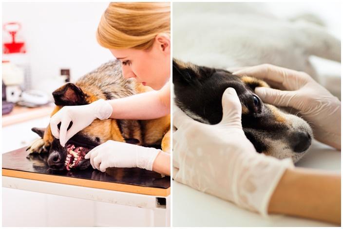
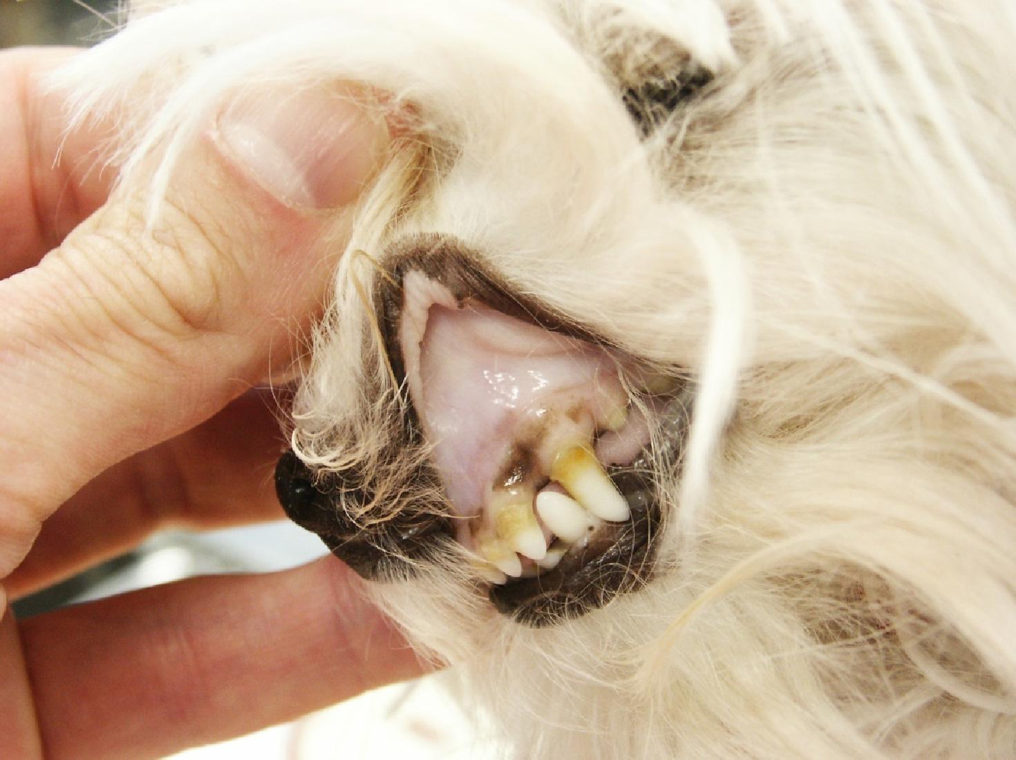
- Gums and oral cavity: we can see the gums by lifting the dog's upper lip and looking into their mouth. It is important to note that some dogs have darker mucous membranes in their mouths due to natural pigmentation. We can still observe changes to their coloration, but they are often more difficult to detect.
- Internal side of the eyelids: we can make an inversion of the eyelid with our thumb, although this should be avoided in case we introduce bacteria to the eye. As with the gums, the inner side of the eyelid can be dark in color.
- Genitals: the glans penis in males can be exteriorized or the vulvar fold in females inverted. If you do not have experience it can be somewhat complicated to do. It can also cause discomfort in the dog due to the sensitive nature of these tissues.
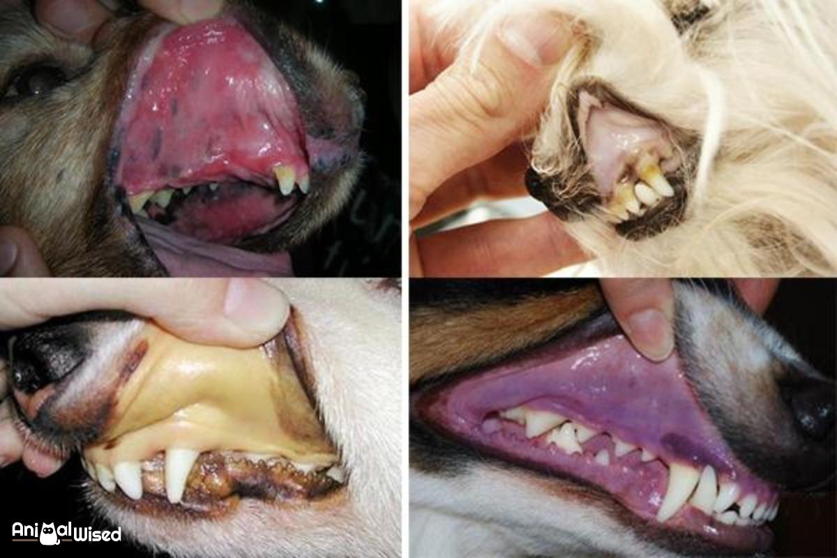
We can know that the mucous membranes of a dog are normal when we observe them as pink, moist and shiny. However, it is important to know that some dogs will have naturally dark mucous membranes in their mouth. This is the case with certain dogs that have naturally black gums such as the Chow Chow or some Shar Pei dogs.
In cases of dogs which have dark gums, the gums won't be pink, but they should still be healthy and vibrant in appearance. When they are ill, they will become paler. For dogs that have naturally pink gums, signs of darkening are usually another indication of illness.
Whatever their natural color, a dog's gums should have a capillary refill time (CRT) of no more than two seconds. Mucous membranes that are dry, tacky or show abnormal CRT suggest an abnormality in the peripheral circulation and may be caused by shock or dehydration of the dog.
Capillary refill time (CRT)
The capillary refill time is the time elapsed between capillaries being drained via compression and then being refilled again. This can be carried out on various parts of the skin. It is easier to perform on humans as we have much of our skin exposed. In dogs, the hair which covers their body can make this difficult. For this reason, the mucous membranes are often the best place to carry out this test since we can see them more easily.
Calculating the capillary refill time is very simple, we only need to press our finger on the mucosa (genital or oral), which will turn white. When we remove our finger, it should return to its normal pink color after two seconds. If the dog is suffering a health problem, the capillary refill time will be longer.
When we see discoloration or paleness in the dog's mucous membranes, the capillary refill time test may take even longer. In extreme cases, there is such an insufficient supply of blood, the CRT test won't work at all. This is because pushing on the mucous membranes will not cause them to become any paler than they already are.
In other cases, we may observe the mucosa are an abnormal color. In this case, we should try another area to see if the problem is localized. In the case of observing mucosa of an abnormal color or a high CRT, it is always advisable to assess a second mucosa. In doing so, we will be able to detect a possible local or generalized issue.
Discover what happens when pathogens cause inflammation of the nasal mucous membranes with our article on the causes and treatment of rhinitis in dogs.

Pale mucous membranes in dogs
When we look at a dog's mucous membranes and see they are pale, it means there is either a decrease in blood flow or in the number of red blood cells present in the blood. It is common in dogs that are in a state of shock, dogs that suffer from internal bleeding or those that are experiencing poisoning. In these cases, the dog is undergoing a medical emergency. If the mucous membranes are completely white, they are in life-threatening trouble.
Pale mucous membranes in dogs are often due to anemia, a disorder of the blood which results in it being unable to transport oxygen. There are many different types of anemia, but they can also result in pale mucous membranes when there are sufficiently low amounts of oxygen in the blood. The level of anemia can be mild or severe, depending on the cause. Pale mucous membranes can present in both mild and severe anemia in dogs.
With this in mind, we look at some of the various reasons why a dog's mucous membranes are pale:
- Hemolytic anemia: this can be acquired, but it is also possibly immune-mediated, meaning it can affect dogs with pre-existing immunity disorders.
- Blood loss: rather than a pathogen affecting oxygen transport, when the dog loses a sufficient amount of blood, it will not longer be able to transport enough oxygen.
- Septic shock: when an infection spread sufficiently, the dog will go into a state of sepsis which threatens their life.
- Dehydration: if the dog is severely dehydrated, it can cause the dog's circulation to become poor and decrease the amount of oxygen being transported.
- Hemophilia: blood diseases can result in poor oxygenation and subsequent poor mucous membranes. Similar blood diseases such as von Willebrand's disease in dogs can cause this problem.
- Low blood pressure: could be due to blood loss, poor kidney functioning, hypothyroidism or other causes.
- Heart problems: if a heart is not functioning properly, it can cause poor oxygenation. This could be due to congenital heart defects, congestive heart disease or other causes of heart failure in dogs.
In an emergency veterinary situation, a vet will look at the mucous membranes of the dog when assessing them. If they are very pale or white, it is likely the dog will be in a serious medical condition.

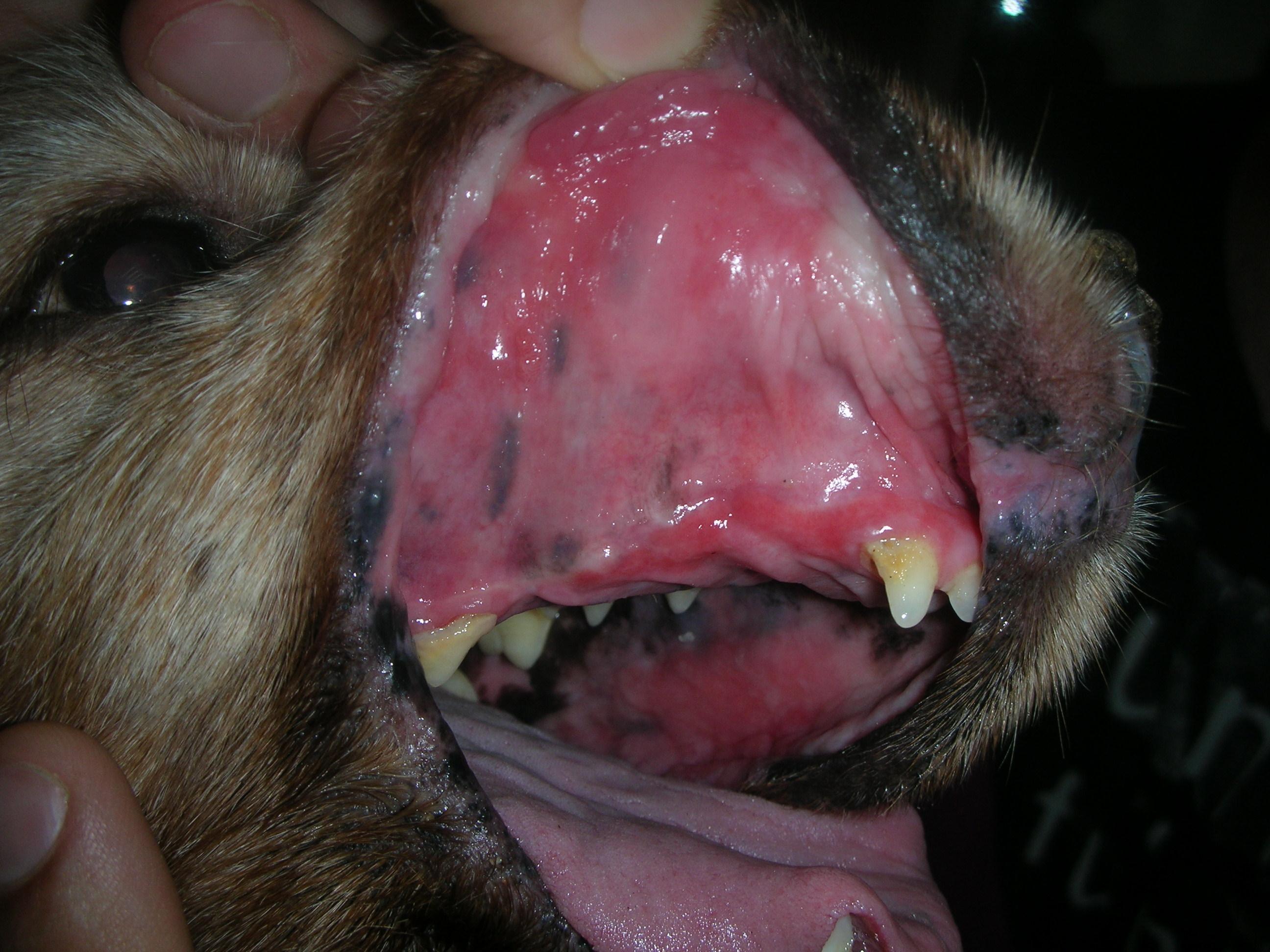
Red mucous membranes in dogs
When we observe the mucous membranes of a dog are of an intense red color, it means they are congested. They indicate an increase in blood flow and the cause of this color may be related to heat stroke in dogs or blood infections such as sepsis. Sepsis is a life-threatening condition and the dog will need to be stabilized in a veterinary clinic.
It is also possibly due to other processes which cause the dog's body to go into shock. These can cause the dog to have brick-red gums due to the high concentration of blood. These can be caused by the following:
- Sepsis: as we have mentioned, sepsis is life threatening in dogs. It is a blood infection which spreads throughout the dog's organism.
- Poisoning: this can be due to the ingestion of toxins, something especially common in food. Dogs are not always discerning with what they eat, sometimes even ingesting toxic chemicals by accident. It can also be due to inhaled toxic gas such as carbon monoxide.
- Heat stroke: being too hot can have a serious affect on the dog and result in them panting, drooling, becoming lethargic and displaying brick-red gums.
- Anaphylactic shock: as a result of an extreme sensitivity to various allergens.
These are all serious problems which can result in the death of the dog. Learn more in our article on sepsis in dogs.

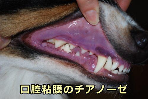
Blue mucous membranes in dogs
We can observe the blue or violet mucous membranes when they have cyanosis. This is an increased concentration of reduced hemoglobin (Hb), indicating a lack of oxygen in the blood. Clinically evident cyanosis typically occurs at an oxygen saturation of 85% or less[1]. It can be caused by the following:
- Suffocation: if the dog simply cannot intake enough oxygen for gas exchange, they will develop cyanosis. This could be due to getting their head trapped in a bag, choking on food or other accidents.
- Respiratory problems: this is a broad term for various problems which can cause breathing difficulties. They include respiratory infections, airway obstruction, lung collapse, pneumonia or pulmonary edema.
- Heart disease: similar to causes of pale mucous membranes in dogs.
- Hypothermia: if the dog is exposed to sufficiently low temperatures, they will go into hypothermic shock. In addition to their mucous membranes, their skin can also go blue.
In addition to the above, ingestion of toxins can cause a dog's gums to turn a bluish color. Learn more with our article on poisoning in dogs.

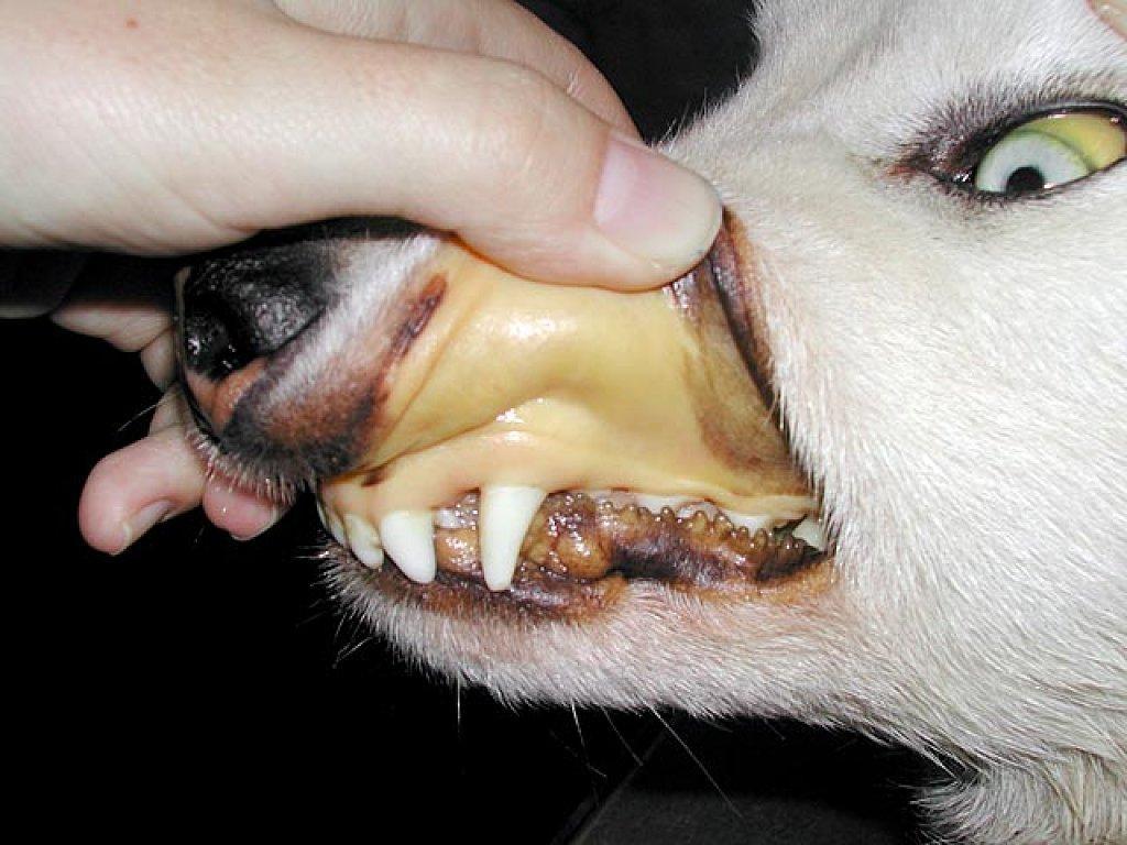
Yellow mucous membranes
We can observe yellow mucous membranes, known as icteric mucosa. It indicates an increase in bilirubin values and can be caused by many causes, ranging from intoxication to hemolysis. This discoloration is often related to liver and kidney functioning. When these organs cannot function, they cannot filter the toxic substances which pass through the body. The result is their accumulation, which can result in jaundice in dogs, amount other issues.
With this in mind, some of the causes of yellow mucous membranes in dogs include the following:
- Jaundice: this is not a disease, but it is a symptom in itself. It can be caused by bacterial infections, intoxication, metabolic disorders and others.
- Hemolytic anemia: can also be due to immune-mediated causes.
- Bile duct obstruction: can be due to stones blocking the duct directly or due to inflammation such as pancreatitis putting pressure on the bile duct.
- Liver diseases: when the liver is no longer able to function, it can lead to a buildup of bilirubin. These include cirrhosis of the liver, tumors or hepatitis.
Other causes include infections, metabolic disorders and other chronic illnesses. Read our related article on why your dog's skin is turning yellow to learn more.

What to do if our dog has mucous membrane discoloration
If we observe congestive or cyanotic or pale mucous membranes, we must go to the vet immediately, since it is a veterinary emergency. However, the other colorations are no less important, we will call our trusted veterinarian to communicate the situation and see what measures to take.
In many cases, the treatment options will be required to slow down the damage caused by different types of liver failure in dogs or other issues. This may mean that some damage has occurred and we need to focus on preventing more damage and managing ongoing symptoms. We will also need to look at any concurrent symptoms to help inform the veterinarian and carry out an accurate diagnosis.
This article is purely informative. AnimalWised does not have the authority to prescribe any veterinary treatment or create a diagnosis. We invite you to take your pet to the veterinarian if they are suffering from any condition or pain.
If you want to read similar articles to Pale Mucous Membranes in Dogs, we recommend you visit our First aid category.
1. Heinzman, D. M. (2007). Chapter 30 - Cyanosis. In L. B. Zaoutis & V. W. Chiang (Eds.), Comprehensive Pediatric Hospital Medicine (pp. 145-148).
https://doi.org/10.1016/B978-032303004-5.50034-X


