Dog Paw Pads Anatomy


See files for Dogs
A dog's extremities play important functions, not only related to mobility. They allow them to interact with their environment, communicate with others and even allow themselves to get comfortable. A dog's paws are almost paradoxical as they are both very sturdy and very sensitive at the same time. Maintaining their paws is important not only for function, but for their health. If they develop any problems in their paws, it will have a consequential effect on other body parts.
At AnimalWised, we help you better understand dog paw pads anatomy. By looking at the front and back paws of a dog we can understand how they help them to maneuver, as well as look at what differences there may be between individuals.
Parts of a dog's paw
A dog's paws are part of their locomotor system which are responsible for supporting the body's weight while upright. Dog paws are made up of the following components:
- Bones: these can be long, short or flat bones. Most bones in a dog's paw are long and designed to act as levers and facilitate movement. Short bones can be found at the level of the carpus or tarsus joints, allowing complex articulated movements. All the leg bones are connected and function with each other. For example, the dog's scapula and pelvis support muscle and protect underlying soft tissues. Dogs have a specific bone known as sesamoid bones. These bones are located within some tendons and serve to prevent their excessive wear.
- Joints: unions of two or more bones to each other. In the extremities of dogs, the vast majority of joints are synovial (wide movement), although there are also some cartilaginous (slight movement) and fibrous (no movement) joints.
- Muscles: through their contraction and relaxation, they allow movement of the limbs. Dogs have 40 muscles in the forelimbs and 36 in the hindlimbs.
- Tendons: bands of connective tissue that connect muscle to bone. They allow the force generated by the muscle to be transmitted to the skeleton so that movement occurs.
- Ligaments: bands of connective tissue that hold joint bones together.
- Blood vessels: arteries carry oxygenated blood to the tissues of the extremities and the veins return the deoxygenated blood back to the heart.
- Lymphatic vessels: carry lymph from the extremities to the lymph nodes where they drain.
- Nerves: structures of the peripheral nervous system that transmit the nerve impulse to the different tissues of the extremities.
- Skin and subcutaneous tissue: act as a physical barrier protecting the underlying tissues of the extremities.
- Other components: keratinized paw pads and nails.
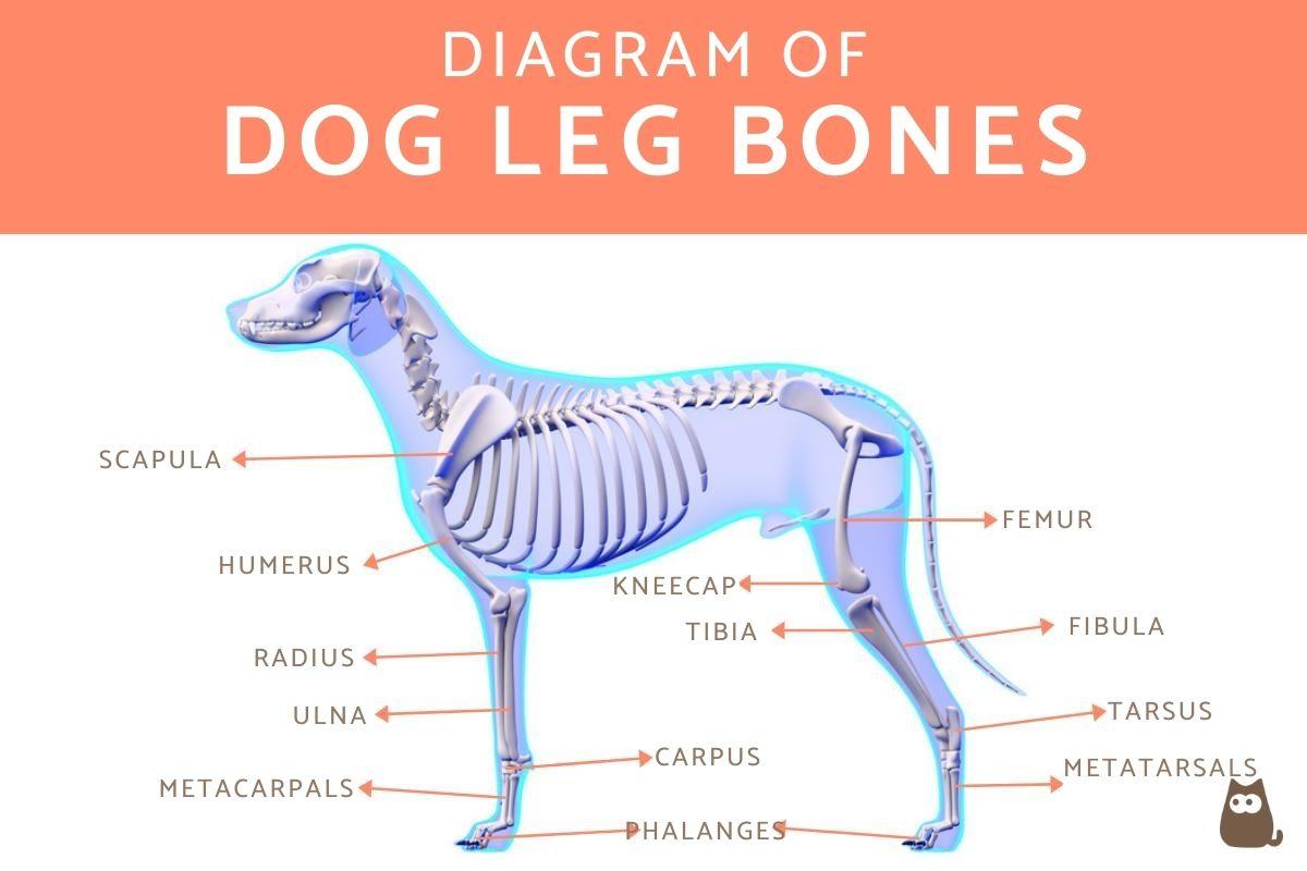
Bones of the forelimbs of dogs
The bones that make up the front legs or forelimbs of dogs are:
- Scapula: it is a flat bone. It should be noted that the scapula remains attached to the leg only by fibrous attachments. Excessive abduction (separation) of the scapula could lead to dislocation and a lesion of the brachial plexus.
- Humerus: long bone that forms the shoulder joint along with the scapula.
- Ulna and radius: two long bones that have an X-shaped spatial arrangement. Together with the humerus, they form the elbow joint. Correspond to the forearm of a human.
- Carpus: made up of two rows of short bones. The proximal row has 3 bones and articulates with the ulna/radius, while the distal row has 4 bones and articulates with the metacarpals. Corresponds to the wrist of humans.
- Metacarpals: there are 5 metacarpal bones and they correspond to the five digits on the dog's paws.
- Phalanges: the first digit has only 2 phalanges, while the remaining four have 3 phalanges (proximal, middle and distal). It should be noted that the proximal and distal sesamoid bones are located at the level of the phalanges.
Bones of the hind limbs of dogs
The bones that make up the hind legs or hindlimbs of dogs are:
- Coxa: is made up of the ilium, ischium and pubis, essentially the dog's hip.
- Femur: a long bone that forms the hip joint together with the.
- Tibia, fibula and patella: the tibia and fibula are two long bones that make up the knee joint (femorotibiorotulian joint) along with the femur and patella.
- Tarsus: it is made up of two rows of bones. The proximal row is made up of 2 bones and the distal row of 4 bones. Corresponds to the human ankle.
- Metatarsals: there are 5 metatarsal bones, but the first is so small that it is at the level of the tarsus (it corresponds to the spur of a human)
. - Phalanges: have the same configuration as in the forelimbs.

Parts of a dog's plantar area
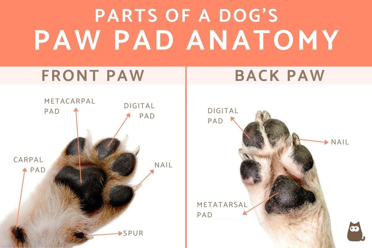
A dog's front paws have 5 digits, sometimes referred to as ‘fingers’. The hind paws only have 4 digits. The exception is when the dog may have one or more dewclaws. You can see this when your dog has 5 toes on their back feet, something caused by genetics and more prevalent in some breeds than others.
We sometimes refer to a dog's front paws as their ‘hands’ and back paws as their ‘feet’. Both have nails known as claws. The paw pads are cushioned structures which make up the plantar area on which the paws touch when they walk. Dogs also have finger pads on their digits.
Each dog paw has soft pads which make up the famous shape we see of a dog paw print. There is a small pad on each digit, the front paw has a metacarpal pad and the back foot has a metatarsal pad, both of which are the largest of their relative paws. The front paw also has a carpal pad located higher than the others. These pads are made up of a collection of fatty tissue covered by thick, dark skin, which in turn is covered by a thick layer of keratin. In puppies, the skin on the pads is softer and finer, but hardens as the dog grows.
If the paws are too rough and hard, it could be due to a condition known as hyperkeratosis in dogs.
The pads serve important functions for the dog:
- They cushion the impact of the extremities against the ground.
- They serve as a thermal insulator.
- They protect the legs from continuous friction with the ground.
- The pads located at the level of the carpus serve to maintain balance on slippery surfaces and help them stop when in motion.
The dog's nails are also made up of keratin and hardened skin cells with living tissue inside. If the nails are light in color, we can see the tissue inside, but this is harder to do if the nails are darker. Dogs generally wear down their nails when walking or running. However, domestication means they can often overgrow which can lead to health problems.
Caring for the plantar area of dogs
In addition to cutting the nails, we must take special care with the paw pads. Despite the fact the pads are very resistant structures, they are subjected to constant abrasion due to their friction against the ground. It is important to take adequate care to maintain condition. We explain the most important care of dog pads:
- Avoid very abrasive surfaces: walking for a long time on very abrasive ground, such as asphalt, cement or beach sand, can wear or even cause erosions or ulceration on the paw pads. To keep your dog's pads healthy, try to stay on softer surfaces such as grass when walking.
- Avoid very hot or very cold ground: in summer, the ground temperature can become very high (especially on dark grounds such as asphalt) and cause burns on your dog's pads. The same can happen on floors covered with ice or snow. For this reason, it is recommended that in summer you take walks in times of less heat and in shady areas. On the contrary, in winter you should look for sunny areas and avoid those with frost or snow.
- Avoid sharp objects: during walks it is important that you pay attention to the presence of sharp or pointed elements (glass, nails, etc.) that can dig into your dog's pads and cause painful ulcers.
- Keep the pads dry: when the pads are immersed for too long in water, they become soft and can be more easily eroded by rubbing against abrasive soil. It is important you dry the pads after your walks, especially if it is raining. Likewise, if you have a dog with a special interest in water, you should control the time of the baths (no more than 15-20 minutes) and make sure they walk on something soft when being dried.
You will need to be very careful with certain seasonal issues. Foxtails are a spiked grass cluster which are sharp enough to embed in your dog's skin, even on their paws. These are more common in summer. We also need to look at other issues such as when the dog breaks a nail.

Curiosities of the legs of dogs
Now that you know the parts of a dog's legs and paws, here are some curious facts you may not know about them:
- Dogs are digitigrade animals, which means they walk with only their toes supported (they do not support the carpal or tarsal joints).
- The morphology of the limbs differs between different breeds of dog. As an example, swimming-adapted breeds such as the Newfoundland or Labrador Retriever have wider bones and longer toes. In contrast, Greyhounds bred for running have longer middle toes.
- Dogs only have sweat glands at the level of the paw pads. This implies they hardly lose heat through the evaporation of sweat and need other mechanisms, such as panting, to regulate their body temperature.
- Some breeds of dogs, such as the Pyrenean Mastiff or the Spanish Mastiff can have a double spur on the hind limbs. It is a vestigial structure that usually does not have any negative consequence, although it needs to be monitored in cases it contributes to any health problems.
- Most of the important limb structures (such as blood vessels, nerves, etc.) are located on the medial side (the side closest to the animal's body), which keeps these structures protected in case of trauma, contusions, bites, etc. It should also be noted that in dogs, especially medium, large and giant breeds, it is common to use the veins of the fore or hind limbs to extract blood samples or place intravenous lines. The cephalic vein is usually used in the forelimbs and the saphenous vein in the posterior limbs.
Not all dogs are the same and some have more sensitive skin than others. This can be due to a genetic predisposition, but it may also be the result of some other issues. Take a look at our article on sensitive dog paws to learn more.
If you want to read similar articles to Dog Paw Pads Anatomy, we recommend you visit our Basic care category.
- Done, SH, Goody, PC, Evans, SA, Stickland, NC (2010). Color Atlas of Veterinary Anatomy . Elsevier.
- McCracken, TO, Kainer, RA (2017). Small Animal Anatomy Atlas. SD Editors.


