Mast Cell Tumor in Cats - Causes of Mastocytoma


See files for Cats
Mastocytoma in cats, otherwise known as mast cell tumors, can be either subcutaneous or visceral. This refers to whether they develop under the skin or on internal tissue, respectively. Subcutaneous mast cell tumors are more common and are the second most prevalent type of malignant tumor in cats. Visceral mast cell tumors most frequently affect the spleen, but can affect other organs. Since they are potentially fatal, we need to be aware of their causes and treatment to best ensure the health of our cats.
At AnimalWised, we present our guide to mast cell tumors in cats. We explain more about the causes of mastocytomas, share how they are diagnosed and tell you what you might be able to expect in terms of treatment.
What is mastocytoma in cats?
Mastocytoma is a tumor that consists of an over-proliferation of mast cells. Mast cells are cells originating in the bone marrow from hematopoietic precursors and can be found in the skin, connective tissue, gastrointestinal tract and respiratory system.
Mast cells are first-line defensive cells against various infectious agents. Their granules contain substances that mediate allergic and inflammatory reactions, such as histamine, TNF-α, IL-6, proteases, etc. They also respond to tissue trauma, so they can help repair wounds and other injuries to tissue.
When a tumor of these cells occurs, the substances contained in their granules are released in an exaggerated way. They cause localized or systemic effects that can give rise to many different clinical signs, depending on their location. Although histamines are important for helping the immune system, if we have too many of them, they can damage the tissue.
Mast cell tumors are relatively common in veterinary medicine. They are known to veterinarians as ‘the great pretenders’ as they can imitate various other types of growths and lesions. Even something which looks like a wart may be a mast cell tumor in disguise.
It is important to understand that these tumors can sometimes be mistaken for benign growths, which complicates diagnosis. Due to their unpredictable nature, regular monitoring and veterinary check-ups are crucial for early detection and treatment.
Source: Assets

Types of mast cell tumor in cats
In cats, the mastocytoma can be cutaneous, when it is located in the skin, or visceral, when it is found in internal viscera. This means they can appear almost anywhere on the cat's body.
Cutaneous mast cell tumor
It is the second most common malignant tumor in cats, and the fourth among all feline tumors. The different types of Siamese cats seem more predisposed to cutaneous mast cell tumors than other cat breeds. There are two forms of cutaneous mast cell tumor according to their histological characteristics:
- Mastocytic: occurs especially in cats over 9 years of age and is divided into a compact form (the most frequent and benign in up to 90% of cases) and a diffuse form (more malignant and most commonly resulting in metastasis).
- Histiocytic: this occurs between the ages of 2 and 10 due to the period of sexual maturity. Interestingly, histiocytic mast cell tumors are often less aggressive, with some resolving spontaneously without intervention.
Visceral mast cell tumor
These mastocytomas can be found in parenchymal organs such as:
- Spleen (most common)
- Small intestine
- Mediastinal lymph nodes
- Mesenteric lymph nodes
Mast cell tumors especially affect older cats between 9 and 13 years of age. This is an important reason why it is important to take senior cats for regular veterinary checkups. Additionally, understanding the risk factors and symptoms can lead to earlier detection and more effective management.
Symptoms of mast cell tumor in cats
Symptoms may vary depending on the type of mast cell tumor in cats. Below we look at the symptoms most commonly associated with subcutaneous and visceral mastocytoma separately.
Symptoms of subcutaneous mast cell tumors in cats
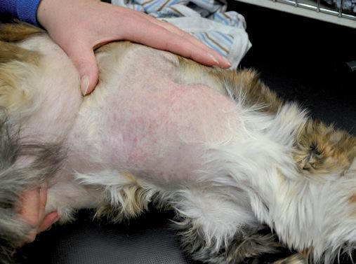
Subcutaneous mastocytomas in the cat can appear as single or multiple neoplasms (the latter occurring in 20% of cases). They can be found on the head, neck, chest or extremities, among other areas.
It consists of nodules that are usually:
- Defined
- 0.5-3 cm in diameter
- Not pigmented or pink
Other clinical signs that may appear in the area of the tumor include:
- Erythema (skin redness)
- Superficial ulceration
- Itching
- Self-trauma
- Inflammation
- Subcutaneous oedema
- Anaphylactic reaction
The nodules of histiocytic mast cell tumors usually disappear spontaneously. Self-trauma occurs when the cat tries to scratch the itching of the mast cell tumor and opens the skin with their claws. Find out other reasons for this in our article on why cats scratch themselves raw.
Symptoms of visceral mast cell tumors in Cats
As you can see, many of the symptoms of subcutaneous mast cell tumors are related to skin problems. Cats with visceral mast cell tumors present with signs of systemic disease such as:
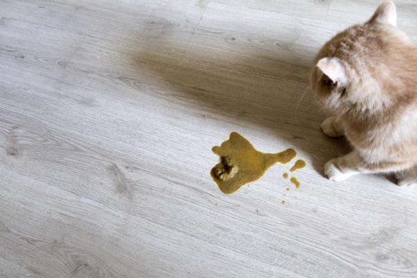
- Vomiting
- Depression
- Anorexia
- Weight loss
- Diarrhea
- Hyporexia
- Respiratory distress (if pleural effusion)
- Splenomegaly (enlarged spleen)
- Ascites
- Hepatomegaly (enlarged liver)
- Anemia (14-70%)
- Mastocytosis (31-100%)
When a cat presents alterations in the spleen, such as an increase in size, nodules or anything affecting the organ, the first suggestion to the veterinarian will be the presence of a mast cell tumor. Early recognition of these systemic signs can lead to prompt treatment and improved outcomes.

Diagnosis of Mastocytoma in Cats
A diagnosis will be made depending on the veterinarian's decision after a general examination. For example, the presence of subcutaneous growths will help suspect a certain type of mastocytoma.
Diagnosis of subcutaneous mastocytoma in cats
Subcutaneous mastocytoma in cats is suspected when a nodule with the characteristics described above appears. It is confirmed by cytology or biopsy. Histiocytic mast cell tumor is the most difficult to diagnose by cytology due to its cellular characteristics, vague granularity and the presence of lymphoid cells.
It must be taken into account that mast cells can also appear in feline eosinophilic granuloma, which can lead to an erroneous diagnosis. Therefore, veterinarians may use additional diagnostic methods to confirm their suspicions and avoid misdiagnosis.
Diagnosis of visceral mast cell tumor in cats
The differential diagnosis of feline visceral mast cell tumor, especially that of the spleen, includes the following processes:
- Splenitis (spleen inflammation)
- Accessory spleen (excess splenetic tissue)
- Hemangiosarcoma (relatively rare sarcoma in cats)
- Nodular hyperplasia
- Lymphoma
- Myeloproliferative disease
The blood count, biochemistry and imaging tests are key to diagnosing visceral mast cell tumor:
- Blood tests: in blood tests, mastocythemia and anemia can lead to suspicion. This is especially with the presence of mastocythemia, an increase of mast cells characteristic of this process in cats. Masyocytemia in cats is almost always due to mast cell tumors, while this is not the case in all other animals[1]. Understanding the unique presentation of these tumors in cats compared to other species is crucial in ensuring an accurate diagnosis.
- Abdominal ultrasound: Abdominal ultrasound can detect splenomegaly or intestinal masses. It will also help to look for metastases in mesenteric lymph nodes or other organs, such as the liver. It also allows us to see alterations in the parenchyma of the spleen or nodules.
- Chest X-ray: a chest X-ray allows us to observe the state of the lungs, looking for metastases, pleural effusion or alterations in the cranial mediastinum. This imaging technique aids in determining the extent of the disease and planning appropriate treatment strategies.
- Cytology: fine-needle aspiration cytology of the spleen or intestine can differentiate a mast cell tumor from other processes described in the differential diagnosis. If performed in pleural or peritoneal fluid, mast cells and eosinophils can be observed.

Treatment of mast cell tumor in cats
The treatment of mast cell tumors in cats will also depend on its location. Some variations depending on the type of mast cell tumor being treated.
Treatment of subcutaneous mast cell tumors in cats
Treatment of cutaneous mast cell tumors is performed with surgical removal. This occurs even in cases of histiocytic forms, which tend to regress spontaneously.
Surgery is curative and must be performed by resection in localized cases and with more aggressive margins in diffuse cases. In general, local excision with margins between 0.5 and 1 cm is suggested for any subcutaneous mast cell tumor diagnosed by cytology or biopsy.
Recurrences in cutaneous mast cell tumors are very rare, even in incomplete excisions. The low recurrence rate further emphasizes the effectiveness of surgical intervention in these cases.
Treatment of visceral mast cell tumor in cats
Surgical removal of visceral mast cell tumors is performed in cats with an intestinal or spleen tumor which has not metastasized. Before removal, the use of antihistamines such as cimetidine or chlorpheniramine is advised to reduce the risk of mast cell degranulation. This can lead to problems such as gastrointestinal ulcers, coagulation abnormalities and hypotension.
The average survival time after splenectomy is between 12 and 19 months, but negative prognostic factors include cats with anorexia, significant weight loss, anemia, mastocytosis (a rare mast cell disorder) and metastasis. It's important for pet owners to understand these factors, as they can influence the overall treatment plan and expected outcomes.
After surgery, complementary chemotherapy with prednisolone, vinblastine or lomustine is usually administered. Chemotherapy helps manage any residual disease and lowers the risk of recurrence.
In cases of metastasis or systemic involvement, prednisolone for cats can be used orally at doses of 4-8 mg/kg every 24-48 hours. If an additional chemotherapeutic agent is needed, chlorambucil can be used orally at a dose of 20 mg/m2 every other week. To improve the symptoms of some cats, antihistamine drugs can be used to reduce excessive gastric acidity, nausea and the risk of gastrointestinal ulcer, antiemetics, appetite stimulants or analgesics. By addressing the symptoms effectively, the quality of life for affected cats can be significantly enhanced.
This article is purely informative. AnimalWised does not have the authority to prescribe any veterinary treatment or create a diagnosis. We invite you to take your pet to the veterinarian if they are suffering from any condition or pain.
If you want to read similar articles to Mast Cell Tumor in Cats - Causes of Mastocytoma, we recommend you visit our Other health problems category.
1. Piviani, M., Walton, R. M., & Patel, R. T. (2013). Significance of mastocytemia in cats. Vet Clin Pathol, 42(1), 4-10.
https://pubmed.ncbi.nlm.nih.gov/23278591/
- Harvey, A., & Tasker, S. (Eds). (2014). Handbook of Feline Medicine. Ed. Sastre Molina, SL L'Hospitalet de Llobregat, Barcelona, Spain.
- Assisi Group. (2020). Feline mast cell tumor. Retrieved from: https://issuu.com/editorialservet/docs/argos_220_mr/s/10700653
- Rios, A. (2008). Canine and feline mastocytoma. Clin Vet Peq Anim, 28(2), 135-142. https://ddd.uab.cat/pub/clivetpeqani/11307064v28n2/11307064v28n2p135.pdf
- Del Castillo, N. (n.d.) Mastocytoma in the cat. Retrieved from: https://www.avepa.org/pdf/vocalias/MTC.pdf