Anatomy of a Frog - Internal and External


Frogs are amphibians, types of vertebrate animals in nature which have the ability to live between water and land. Frogs are perhaps the most emblematic of these animals, but there is also great diversity among the many frog species we can find. All of them have certain physical and behavioral characteristics which they share. While we may think of them as docile and even rather cute, frogs are largely carnivorous and some are deadly poisonous. In order to carry out their various functions, they require a physiology which is specifically adapted.
At AnimalWised, we learn more about how frogs are uniquely adapted to their environments by looking at both the internal and external frog anatomy. We do so before and after their metamorphosis, meaning we show both tadpoles and adult frogs. We also provide diagrams so you can have a better idea of what they look like.
Anatomy of a tadpole
Frogs and toads are from the order Anura. While we rightly associate tadpoles with anurans, not all frog species have a tadpole stage[1]. They all have a larval stage and require water to develop. Frogs that don't have a tadpole stage go through direct development. This means they emerge from the egg as a froglet, i.e. a miniature version of their adult frog stage. This occurs in a minority of frog species as most larval stages are tadpoles.
Tadpoles have a tail which allows them to swim in water, an appendage which is not carried over into their adult stage. Tadpoles have other anatomical structures which allow them to survive in water including gills. They lose these structures due to the process of metamorphosis. Animals that go through metamorphosis change significantly from larval to adult stages.
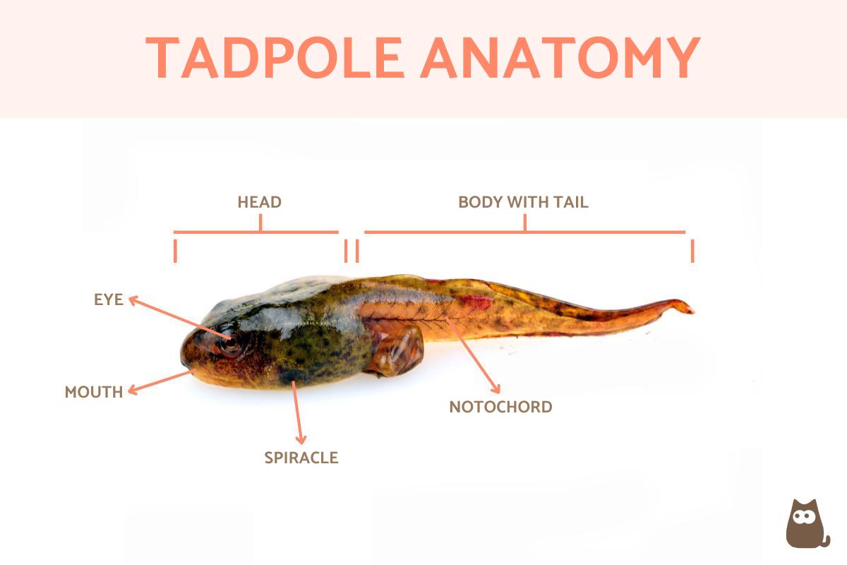
We now look at some of the most important internal and external anatomical features of tadpoles:
- Head: as shown in the diagram below, you can see the head is significantly differentiated from the body. Although they lack eyelids, tadpoles have eyes which are located towards the front of the head. This positioning allows them to have a wider field of vision, which is crucial for detecting predators and navigating their aquatic environment.
- Mouth: they do not have true teeth, but tadpoles have similar structures made of keratin that are located in the upper jaw used for feeding. They may also have these structures in the lower jaw surrounded by a kind of horny beak, but this can vary between frog species. Larvae usually follow a herbivorous diet, but in certain circumstances they can be omnivorous, even eating other tadpoles. Learn more with our article on what tadpoles eat.
- Body: it is elongated. Tadpoles are exclusively aquatic in habit. For this reason, their head is differentiated from an elongated body which ends in a tail. If they resemble fish it is precisely because this body shape allows them to swim in water. They do not have other limbs since they do not need them until after they undergo metamorphosis. Interestingly, the streamlined body also aids in reducing water resistance, enabling efficient movement.
- Tail: a hardened muscular structure that covers the notochord, a cartilage-like structure which is important for sending signals during development. It is an anatomical structure common to all chordates, giving rise to their nomenclature. The tissue of the tail is rigid, but it must be able to decompose during metamorphosis. This occurs due to apoptosis, a type of programmed cell death. Tadpoles are able to shed their tails in some circumstances, such as when attacked by predators. This ability is known as autotomy and is a defense mechanism that allows them to escape.
- Belly: tadpoles have a bulging belly which is part of their digestive system. This includes a digestive tube which starts at the mouth and connects to the intestine. Another tube located towards the end of the tail known as the ventral tube is used for excretion. The digestive system is adapted to process plant material efficiently, ensuring the tadpole gets the necessary nutrients for growth.
- Skeleton: is of a cartilaginous type, but these structures will also transform until bones are formed in adult individuals. As usually occurs in vertebrates, the notochord is transitory (as we have commented) and gives rise to the vertebral column, also ossified into bone in frogs. This transformation from cartilage to bone is crucial for supporting the frog's body on land.
- Respiratory system: they have external gills for breathing. As the tadpole develops, internal gills appear below the initial external ones and are covered by a fold of tissue. Once formed, they create tiny openings to the outside known as spiracles. The tadpole uses the internal gills for gas exchange, but the adult frog does not. Similarly, tadpoles have two nostrils that will later be the nasal openings in the adult. The transition from gills to lungs is a remarkable adaptation that allows frogs to thrive both in water and on land.
As summation, the anatomical structures of a tadpole are as follows:
- Head
- Eyes
- Nostrils
- Oral disk or mouth
- Keratin dental structures
- Spiracle
- Digestive tube
- Cartilaginous skeleton
- Tail
- Cloaca

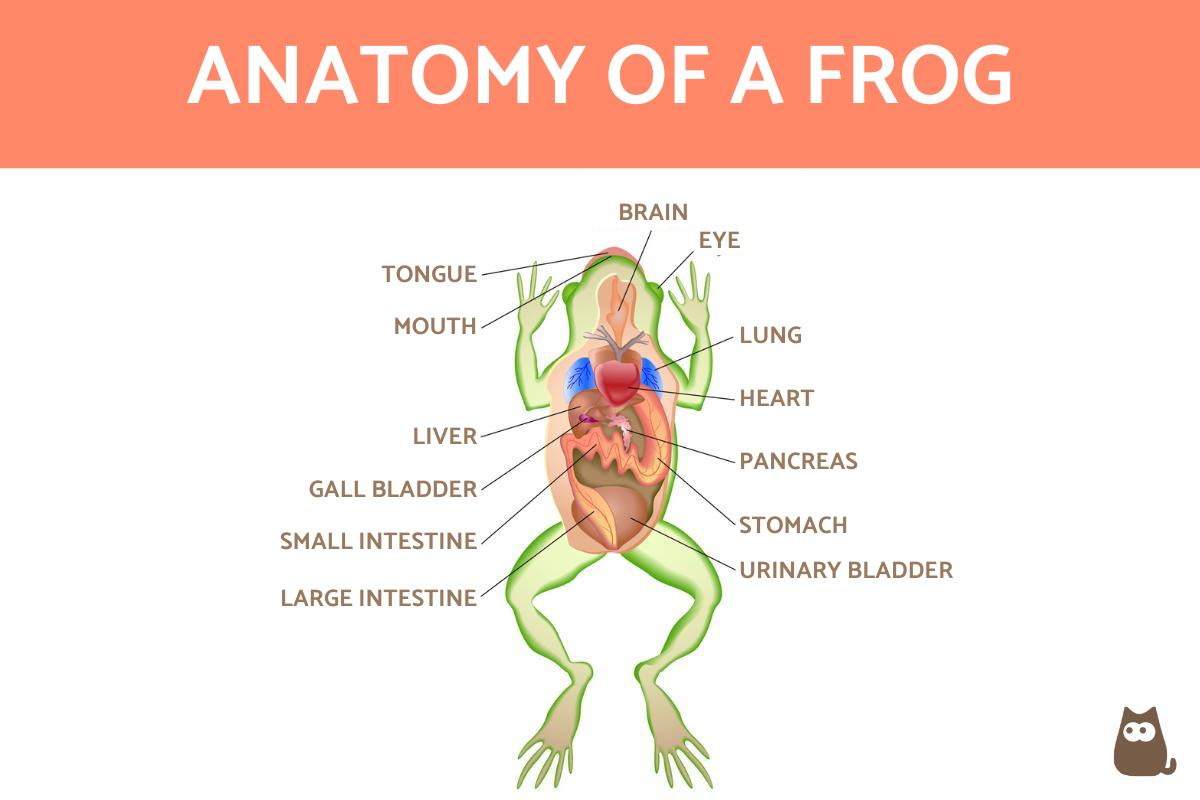
Internal anatomy of a frog
Once metamorphosis occurs, some tadpole structures are transformed or lost when they become adult. The reason for this is mainly due to their habits, since adult amphibians can live on both land and water. For a frog to have both aquatic and terrestrial habits, they require various internal parts of their anatomy. We explain what they are, as well as show their location in the diagram below.
Musculoskeletal system
Frogs are vertebrate animals, so once fully developed they have muscles and a bone structure. Despite their size, this structure is complex. They have around 30 bones which are also present in other vertebrates. However, several of a frog's bones and muscles have been modified to allow them their supreme jumping ability. This is because they move in a saltatory way, leaping from one spot to another rather than moving gradually. Additionally, their strong hind legs and flexible spine provide them with the necessary power and agility for such movements.
Depending on the species, there may be variations in musculoskeletal structure. For example, some frog species are arboreal and have forelimbs which are adapted to provide a specific dexterity and grasping ability[2]. Learn more about the musculoskeletal system of these animals in our related article on whether frogs have bones.
Nervous system
The nervous system of frogs consists of the following main structures:
- Brain
- Spinal cord
- Cranial and spinal nerves
The frog's brain has olfactory lobes, two hemispheres, the pineal body, the optic lobes and the cerebellum, as well as the medulla oblongata. They are responsible for controlling the various senses of the frog which make them both able to predate on other animals, as well as avoid being prey themselves. The brain's structure allows for quick reflexes and responses to environmental stimuli, aiding in survival.
Respiratory system
The respiratory system of frogs includes lungs, organs present in adult frogs, but not in their larval stages. In this way, we can say that frogs have pulmonary respiration. Frogs use their mouth to pump air through their respiratory system into their lungs. Moreover, the ability to absorb oxygen through their skin helps them remain submerged for extended periods without surfacing for air.
Frogs are also animals that can breathe through their skin. This is because their skin is semi-permeable, something especially useful for frogs living in mainly aquatic habitats. This is because these habitats can be deficient in oxygen. The skin's permeability is maintained through a constant layer of mucus, ensuring optimal gas exchange.
Circulatory system
Like other amphibians, frogs have a heart with three chambers through which blood passes to be oxygenated. It does do so by entering through separate atria. These chambers contract to move blood to the ventricle, then pass through a valve and reach the aorta if it is oxygenated blood or the pulmonary artery if it is deoxygenated. This unique circulatory setup allows for both pulmonary and cutaneous respiration, optimizing oxygen uptake.
Digestive system
The frog's digestive system begins in the mouth. Frogs have some teeth that are used to hold prey, but not for chewing. They also have a tongue which is especially adapted to catch prey. It is extendable and has a process of adhesion which is not well understood, but it is believed to be due to a distinct tongue musculature and specially adapted mucus fibrils[3]. The rapid extension of the tongue is a marvel of evolutionary adaptation, allowing frogs to snatch prey with precision.
Once captured, the food moves through the esophagus to the stomach. It is then mixed with digestive enzymes to pass into the small intestine and continue digestion. Within this system, frogs also have a pancreas, large intestine, gallbladder, spleen and its own vascular system. Learn more about frog diet with our article on what do frogs eat?
Excretory system
The parts of the excretory system of frogs are made up of the kidneys, ureters, bladder and cloaca. All these organs are involved in the storage, transport and expulsion from the body of all those waste substances produced by metabolic processes. The cloaca is the orifice for the reproductive, digestive and urinary systems. This multifunctional opening is a characteristic feature of many amphibians and reptiles, simplifying bodily waste expulsion.
Reproductive system
Frogs do not have all the reproductive organs common to many other vertebrates. In the case of males, they have two testicles associated with the kidneys, efferent ducts and urogenital ducts. Females have ovaries also linked to the kidneys and oviducts. It's fascinating to note that many frog species engage in vocalizations to attract mates, a crucial part of their reproductive strategy.
Learn how these processes are carried out with our article on frog reproduction.
Visual and auditory system
Frogs have good binocular vision which is why they have internal nerves associated with the visual system. Although they don't have ears, they do have eardrums, as well as middle and inner ears. This means frogs are able to hear. Although we do not know the extent of their auditory ability, there is some evidence to suggest certain frog species have ultrasonic communication abilities[4]. This advanced auditory capability aids in communication, especially in dense, noisy environments like rainforests.

External anatomy of a frog
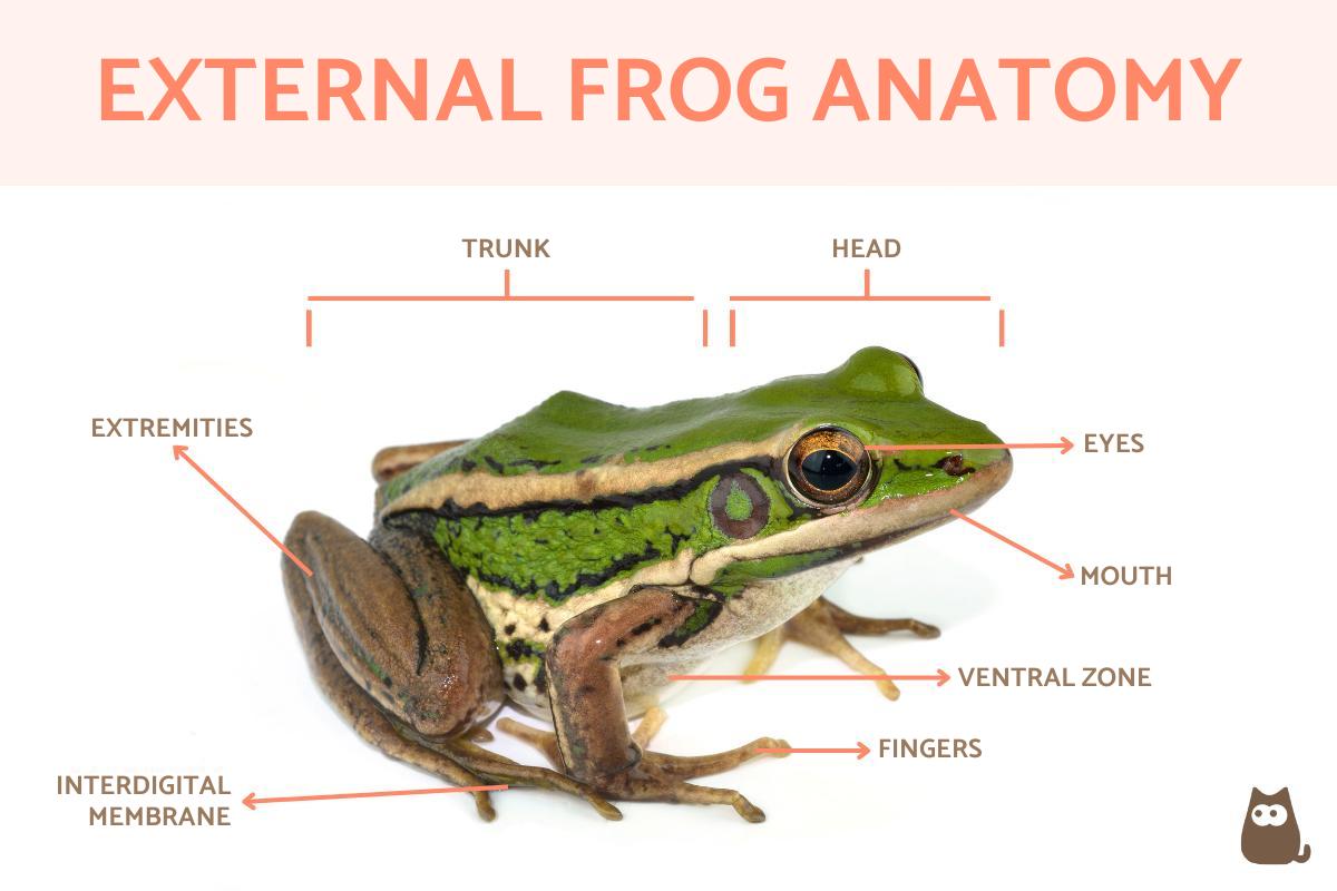
Regarding the external parts of adult frog anatomy, we can find the following important structures which are also shown in the diagram below:
- Head: where the eyes and nasal cavities are located. The positioning of the eyes provides a wide field of vision, crucial for spotting both prey and predators in their environment.
- Trunk: dorsal zone which houses the vital organs.
- Ventral area: the underside or belly. The skin here is often softer and plays a role in respiration, especially in water-dwelling species.
- Limbs: two forelimbs, usually four-fingered, and two five-fingered hindlimbs. The hindlimbs are particularly powerful, enabling the frog's characteristic jumping ability.
The shape of frog legs often varies between species, but the presence of a web between the toes of the hind feet is common. This webbing is an adaptation that enhances swimming efficiency, allowing frogs to propel themselves swiftly through water. The external skin of frogs can be of varying colors, with some being drably colored and others vividly so. Some frogs even have transparent skin which allows us to see their internal organs. This transparency can serve as camouflage, helping them blend into their surroundings.
For a long time frogs have been used in schools and other centers of learning to teach about animal anatomy via dissection. With the advancement of technology, we are able to use didactic models to view both the internal and external parts of a frog's anatomy without having to kill animals for study. These models offer a humane alternative, providing detailed insights into their complex anatomy.
Learn more about these incredible amphibians by looking at our sister site's guide to the difference between toads and frogs.


If you want to read similar articles to Anatomy of a Frog - Internal and External, we recommend you visit our Facts about the animal kingdom category.
1. Gomez-Mestre, I., Pyron, R. A., & Wiens, J. J. (2012). Phylogenetic analyses reveal unexpected patterns in the evolution of reproductive modes in frogs. Evolution; international journal of organic evolution, 66(12), 3687–3700.
https://doi.org/10.1111/j.1558-5646.2012.01715.x
2. Manzano, A. S., Abdala, V., & Herrel, A. (2008). Morphology and function of the forelimb in arboreal frogs: specializations for grasping ability?. Journal of anatomy, 213(3), 296–307.
https://doi.org/10.1111/j.1469-7580.2008.00929.x
3. Kleinteich, T., & Gorb, S. N. (2015). Frog tongue acts as muscle-powered adhesive tape. Royal Society open science, 2(9), 150333.
https://doi.org/10.1098/rsos.150333
4. Feng, A. S., Narins, P. M., Xu, C. H., Lin, W. Y., Yu, Z. L., Qiu, Q., Xu, Z. M., & Shen, J. X. (2006). Ultrasonic communication in frogs. Nature, 440(7082), 333–336.
https://doi.org/10.1038/nature04416
- Hickman, C., Roberts, L., & Parson A. (2000). Comprehensive principles of zoology. McGraw Hill Inter-American: Spain.
- Mijares-Urrutia, A. (1998). The tadpoles of the high Andean anurans (Amphibia) of Venezuela: external morphology and keys. Journal of Tropical Biology, 46 (1), 119-143.
https://revistas.ucr.ac.cr/index.php/rbt/article/view/19360
- Urbina-Cardona, N., Ramirez Pinilla, M. P., & Cortés-Gómez, A. (2016). Functional trait measurement protocol in amphibians.
https://www.researchgate.net/project/MONITORING-FUNCTIONAL-TRAITS-HANDBOOK-Conceptual-and-methodological-protocols-and-applications


